
Ovarian Tumours - Types, Symptoms, Diagnosis And Homeopathic Medicines!


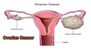

Ovarian tumours are classified as non-cancerous (benign) and cancerous tumours. The non- cancerous tumours also include cysts. The non-cancerous tumour usually do not cause pain but some of them may cause pain. Non-cancerous tumour grow slowly while the cancerous tumours grow very fast and pain.
TYPES OF OVARIAN TUMOURS-
* Non- Cancerous (benign) ovarian tumors
* Cancerous ovarian tumors
NON- CANCEROUS OVARIAN TUMOURS
* Benign cystic teratomas (dermoid cysts)
These tumors usually develop from all three layers of tissue in the embryo (called germ cell layers). All organs form from these tissues. Thus, teratomas may contain tissues from other structures, such as nerve, glandular, and skin tissues.
* Fibromas
These tumors are solid masses composed of connective tissue. Fibromas are slow-growing and are usually less than 3 inches in diameter. They usually occur on only one side.
• Cystadenomas-
These fluid-filled cysts develop from the surface of the ovary and contain some tissue from glands in the ovaries.
SYMPTOMS OF NON- CANCEROUS OVARIAN TUMOURS-
Most functional cysts and noncancerous ovarian tumors do not cause any symptoms. Occasionally, the pelvic area aches or pain occurs during sexual intercourse.
Other Symptoms Are:
* Vaginal bleeding
* Copious menstruation
* If corpus luteum cysts bleed, they can cause pain or tenderness in the pelvic area
* It may be fever, nausea, and vomiting
* Occasionally, sudden, severe abdominal pain occurs because of a large cyst or mass
* Accumulation of fluid in the abdomen called ascitis may occur with fibromas and ovarian cancer
DIAGNOSIS OF NON- CANCEROUS OVARIAN TUMOURS
* A pelvic examination
* Usual Ultrasonography
* Transvaginal Ultrasonography
* If the diagnosis is still unclear, magnetic resonance imaging (MRI) or computed tomography (CT) may be done. If these tests suggest that the growth could be cancerous, doctors remove it and examine it under a microscope. A laproscope is inserted through a small incision just below the navel, may be used to examine the ovaries and to remove the growth.
* Doctors may also do blood tests to check for substances called tumor markers, which may appear in the blood or may increase when some cancers are present. These tests can help confirm or rule out cancer.
CANCEROUS OVARIAN TUMOURS
* Epithelial tumors-
About 90 percent of ovarian cancers develop in the epithelium, the thin layer of tissue that covers the ovaries. This form of ovarian cancer generally occurs in postmenopausal women.
* Germ cell carcinoma-
Germ cell carcinoma can occur in women of any age, it tends to be found most often in women in their early 20s. Six main kinds of germ cell carcinoma exist, but the three most common types are:
- Teratoma
A teratoma is a tumor made up of several different types of tissues including hair, muscle and bone. They typically form in the ovaries, testicles and tailbone and less commonly in other areas.
- Dysgerminoma
A synonym of germinoma is "Dysgerminoma." That
usually occurs in the ovary in adolescence and early adult life. About 5% occurs in pre-pubertal children. Dysgerminoma is extremely rare after age 50. Dysgerminoma occurs in both ovaries in 10% of patients.
- Endodermal sinus tumors (yolk sac tumor)
They occur in the pineal region, ovaries and testicles.
* Stromal carcinoma
Ovarian stromal carcinoma accounts for about five percent of ovarian cancer cases. Unlike epithelial ovarian carcinoma, 70 percent of stromal carcinoma cases are diagnosed in Stage I. They develop in the connective tissue cells that hold the ovary together. The two most common types are:
- Granulosa cell tumor
Granulosa cell tumors are rare ovarian tumors in the stromal cell group.
- Sertoli-Leydig cell tumors
The Sertoli Leydig cell tumors are a group of tumors composed of Sertoli cells, Leydig cells, and poorly differentiated neoplasms. Sertoli–Leydig cell tumor is a member of the sex cord-stromal tumor group of the ovarian and testicular cancer. The tumor can occur at any age and is rare and occurs most often in young adults.
* Small cell carcinoma
Small cell carcinoma of the ovary (SCCO) is a rare, highly malignant tumor that affects mainly young women, with a median age of 24. The subtypes of SCCO include:
- pulmonary small cell carcinoma
- neuro-endocrine small cell carcinoma
- hypercalcemic small cell carcinoma
SCCO accounts for 0.1 percent of ovarian cancer cases. Approximately two-thirds of patients with SCCO have hypercalcemia. The symptoms are the same as other types of ovarian cancer.
SYMPTOMS OF CANCEROUS OVARIAN TUMOURS
Early staged ovarian cancer can produce these symptoms.
* Bloating
* Pelvic or abdominal pain
* Difficulty eating
* Urinary symptoms (urgency or frequency)
Other Symptoms are:
* fatigue
* pain with intercourse
* menstrual irregularities
However, these other symptoms are not so useful in identifying ovarian cancer as the real once.
SYMPTOMS OF SERTOLI- LEYDIG CELL TUMOUR
Due to excess testosterone secreted by the tumor, one-third of female patients present with a recent history of progressive masculinization which is preceded by anovulation, oligomenorrhoea, amenorrhoea and defeminization.
Additional signs include acne and hirsutism, voice deepening, clitoromegaly, temporal hair recession, and an increase in musculature. Serum testosterone level is high.
SYMPTOMS OF GRANULOSA CELL TUMOUR
Granulosa cell tumors are rare ovarian tumors in the stromal cell group that presents with the following symptoms:
Postmenopausal Women:
* Diagnosis of endometrial hyperplasia (thickening of the uterus that causes bleeding) or cancer
* Breast tenderness
* Vaginal secretions
* Virilizing symptoms due to increased testosterone (When a woman starts to show male pattern traits like facial hair growth, for example.)
Premenopausal Women:
* Increased abdominal girth
* Enlarging abdominal mass
* Period Irregularities
Prepubertal Girls:
* Early onset puberty (70-80%) with early male trait characteristics
* Virilizing symptoms due to increased testosterone (When a woman starts to show male pattern traits like facial hair growth)
DETECTION & DIAGNOSIS OF CANCEROUS OVARIAN TUMOURS
* Early Detection
Early detection of ovarian cancer saves women’s lives. No screening test exists except mammograms for breast cancer and Pap tests for cervical cancer.
* Blood Test: CA-125
CA-125 is a protein in the blood that may increase during cancerous tumor. The protein is produced by ovarian cancer cells and is elevated in more than 80 percent of women with advanced ovarian cancers and in 50 percent of those with early-stage cancers.
CA-125, however, is approved by the Food and Drug Administration to monitor the effectiveness of treatment for ovarian cancer and for detecting disease recurrence after treatment.
Although the CA-125 blood test is more accurate in postmenopausal women, it is not a reliable early detection test for ovarian cancer. In about 20 percent of advanced stage ovarian cancer cases and 50 percent of early stage cases, the CA-125 is not elevated even though ovarian cancer is present. As a result, doctors generally use the CA-125 blood test in combination with a transvaginal ultrasound. Because CA-125 misses half of the early cancers and can be elevated by benign conditions, the National Cancer Institute does not endorse using it to screen women for ovarian cancer who are at ordinary risk or in the general population.
* Blood Test: OVA-1
OVA1 has also been approved by the Food and Drug Administration (FDA). A woman who presents with a known tumor may have this test to determine if her surgery should be done by a gynecologist or gynecologic oncologic doctors.
The test measures the levels of five proteins in the blood that change when ovarian cancer is present. However, this test has not been approved for use as an ovarian cancer screening tool, nor has it been proven to result in early detection or reduce the risk of death from this disease.
* Blood Test: Inhibin B and Inhibin A
Granulosa cell tumors are most often detected and monitored via the following blood indicators Inhibin B and Inhibin A.
* Transvaginal Ultrasound-
A transvaginal ultrasound is a test used to examine a woman’s reproductive organs and bladder and can often reveal if there are masses or irregularities on the surface of the ovaries and within cysts that form within the ovaries. To perform the test, the doctor inserts a probe into the woman’s vagina. The probe sends off sound waves which reflect off body structures. The waves are then received by a computer that turns them into a picture.
* CT scan or computerized tomography-
CT scans employ x-rays to take multiple cross-sectional images of the tissues and bones in the body. Doctors can analyze the images individually or use software to make a three-dimensional model of the internal organs. CT scans help define the boundaries of a cancerous tumor and show the extent of tumor spread, helping a doctor determine where to operate. CT scans also are used to monitor disease recurrence. Before undergoing a CT scan, you may receive by mouth or intravenously a contrast material that allows tissues and organs to show up more readily.
* Pelvic Exam-
A pelvic exam may be included as part of a woman’s regular female health exam. This exam requires the doctor to place one or two fingers into a woman’s vagina and another over her abdomen to feel the size, shape and position of the ovaries and uterus. Ovarian cancer is rarely detected in a pelvic exam and usually in an advanced stage if it is.
* Recto-vaginal Pelvic Examination (also called a bimanual exam)-
This exam allows your doctor to examine the ovaries for lumps or changes in shape or size. Every woman should undergo a rectal and vaginal pelvic examination at her annual check-up with her gynecologist. A Pap test is routine in a pelvic exam but it detects cervical cancer, not ovarian cancer
* The need for a biopsy
None of the above tests are definitive when used on their own. They are most effective when used in combination with each other. The only way to confirm the presence of ovarian cancer suspected by the tests is through a surgical biopsy of the tumor tissue.
A doctor may perform laparoscopic surgery to perform the biopsy and remove a small, benign cyst or early ovarian cancer and to determine the extent of spread. A laparoscope is a thin tube with a camera that allows the doctor to see and remove tissue. If a woman has fluid inside the abdomen, a doctor before surgery may inject a needle through the abdomen wall to collect the fluid for analysis. By looking at the cells in the tissue and fluid under a microscope, a pathologist describes cancer as Grade 1, 2, or 3. Grade 1 is most like ovarian tissue while Grade 3 cells are more immature and more likely to metastasize.


+1.svg)
