

Oculomotor nerve (Human Anatomy): Image, Functions, Diseases and Treatments
Last Updated: Mar 18, 2023
Oculomotor Nerve Image
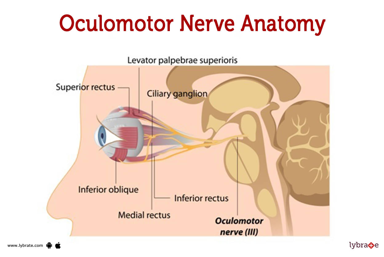
The nerve which is having origin from the brain directly and classified third in number is called oculomotor nerve . It allows eye movements like focusing on a moving object. Your eyes can be moved up, down, and side to side thanks to the third branch of the cranial nerve.Cranial nerve III coordinates its activities with those of the other cranial nerves to provide sensory feedback and to govern eye movement.The olfactory nerve regulates smell (CN I). The optic nerve is in charge of vision (CN II). The trigeminal nerve (CN V) is responsible for facial sensation. Balance and hearing are enabled through the vestibular and cochlear nerves (CN VII).
What is the anatomy of cranial nerve III?
- The Cranial nerve III circuit begins in the middle of the brain.. It travels through a number of structures in the head before reaching the back of the eyes. Its path includes
- Exiting the anterior midbrain.
- Passing by adjacent arteries.
- Penetration of the brain's thick outer layer (dura).
- The cavernous sinus can be accessed through the back of the nose (a hollow space).
- Exiting the skull via the orbital fissure, a big circular opening behind each eye.
- Connecting to the eye's back part.
- Separating into branches that are superior and inferior.
- Both the superior and the inferior branches are connected to the four muscles that control the movement of the eye. They also connect to a muscle in the upper eyelid and muscles inside the eye that control the size of the pupil and how the lens is focused.
Among these are:
- Inferior oblique, which regulates how the eyes turn, how high you look, and how far out you look.
- Inferior rectus, which is in charge of looking down.
- The medial rectus controls the way you look inward.
- Superior rectus, which is in charge of looking up.
- Levator palpebrae superioris is the muscle that determines whether or not you are able to lift the upper eyelid of your eye.
Oculomotor nerve Functions
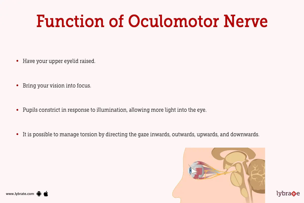
It regulates 4 of the 6 muscles that allow for eye movement. As a result of CN III, you can now do the following:
- Have your upper eyelid raised.
- Bring your vision into focus.
- Pupils constrict in response to illumination, allowing more light into the eye.
- It is possible to manage torsion by directing the gaze inwards, outwards, upwards, and downwards.
How does CN III work?
The following actions are coordinated with eye movement:
- The ability to focus on a target that is either approaching or receding from the observer, known as 'accommodation.'
- It's an optokinetic reflex, where your eyes automatically shift back to their original position after you've finished focusing on an object.
- Using rapid eye movements called saccades, you can move your focus rapidly from one thing to another.
- Maintaining focus on a moving target requires a visual tracking ability known as smooth pursuit.
- The vestibular-ocular reflex is responsible for keeping one's eyes in the proper position while moving one's head.
.jpg)
Oculomotor nerve Conditions and Disorders
Oculomotor dysfunction can be caused by a variety of conditions, including the following:
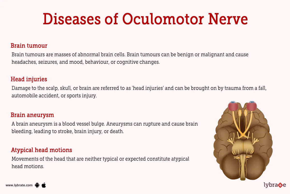
- Brain aneurysm: A brain aneurysm is a blood vessel bulge. Aneurysms can rupture and cause brain bleeding, leading to stroke, brain injury, or death. Brain aneurysms may trigger a sudden terrible headache, dual vision, or a lack of consciousness.
- Brain tumour: Brain tumours are masses of abnormal brain cells. Brain tumours can be benign or malignant and cause headaches, seizures, and mood, behaviour, or cognitive changes.
- Head injuries: Damage to the scalp, skull, or brain are referred to as 'head injuries' and can be brought on by trauma from a fall, automobile accident, or sports injury. From minor (concussion) to serious (traumatic brain injury), head injuries can result in a variety of symptoms, such as headache, dizziness, memory issues, and loss of consciousness.
- Demyelinating disease (multiple sclerosis): Multiple sclerosis is a demyelinating illness that destroys the myelin sheath that protects nerve fibres in the brain and spinal cord. Weakness, muscle spasms, problems, issues with sensation and sight are just a few of the signs and symptoms of demyelination.
- Microvascular disease (diabetes and high blood pressure): Diabetes and high blood pressure are examples of microvascular illness, which is a phrase used to describe conditions that affect the body's small blood vessels, which include those in the brain and eyes. Diabetes and high blood pressure are two conditions that can harm blood vessels and reduce blood flow, resulting in symptoms like eyesight loss, stroke, or organ damage.
- Migraine: Migraine is a form of headache characterised by extreme pain, usually on one side of the head, and may be accompanied by uneasiness, vomiting, and light and sound sensitivities. Migraines can be provoked by a variety of circumstances, and can be controlled with medicines and other treatments.
- Transtentorial brain herniation: A condition in which a portion of the brain is forced out of its usual position by an increase in tension inside the skull. This can lead to side effects such as nausea, vomiting, and shifts in consciousness.
- Third Nerve Palsy (Oculomotor Nerve Palsy): Third Nerve Palsy (Oculomotor Nerve Palsy) is a disorder characterised by damage or compression of the third cranial nerve, which controls many muscles that move the eye. This can result in problems such as double vision, blurred vision, crossed eyes, and abnormal head movements.
- Atypical head motions: Movements of the head that are neither typical or expected constitute atypical head motions. These can include abnormal or exaggerated head motions, as well as trouble controlling head movements.oculomotor nerve Tests
Oculomotor Nerve Tests
- Clinical evaluation: Clinical evaluation is a sort of medical examination done by a medical professional to examine the health status of a patient and diagnose any medical issues. Typically, a clinical evaluation includes obtaining a medical history, doing a physical exam, and prescribing any necessary tests.
- CT: Computed tomography (CT) is a type of diagnostic imaging test which uses X-rays and computers to make detailed pictures of the body's tissues and organs. CT scans can be used to find out if someone has a tumour, an infection, or an injury, among other things.
- MRI: for oculomotor disorders, an MRI may be ordered to assess for any underlying problems with the eyes or surrounding structures that could be contributing to the disorder. It can provide important information to aid in the diagnosis and treatment of these conditions.
- Tonometry: Tonometry is a test used to find out how much pressure is in the eye (intraocular pressure). This test is necessary because glaucoma, a disease that can cause vision loss, can be caused by high intraocular pressure.
- Slit lamp examination: A slit lamp examination is a procedure in which the cornea, iris, and lids of the eye are examined through a magnifying lens. A wide range of medical issues, from infections to injuries, can be identified using this test.
- Fundoscopic exam: A fundoscope is a diagnostic tool used in a fundoscopic exam to observe the retina and optic nerve at the back of the eye.
- Visual acuity test: A test known as a visual acuity test is one that measures how clearly a person can see objects that are close up. This test, which is used to diagnose vision disorders like myopia, farsightedness, and astigmatism among others, often involves reading letters of varying sizes that are displayed on a chart.
- Fluorescein angiography: Fluorescein angiography is a sort of medical imaging test that makes use of a dye to reveal the blood vessels in the eye. This test is capable of diagnosing a wide range of diseases, such as retinal detachment, macular degeneration, and some other issues as well.
Oculomotor Nerve Treatments
- Surgery to relieve pressure on the nerve: Surgery to reduce nerve pressure removes obstructions or damage from the oculomotor nerve. This may involve removing a tumour or blood vessel damage. Surgery relieves nerve pressure and improves symptoms.
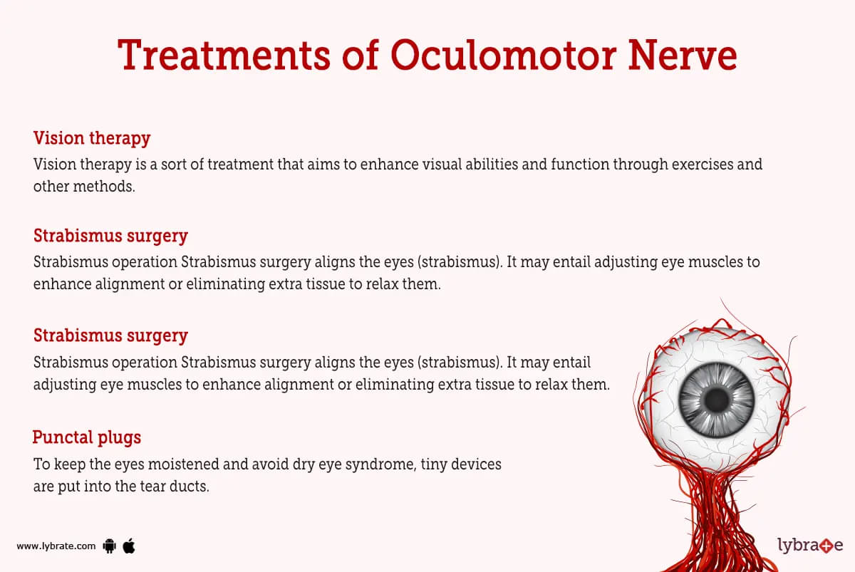
- Strabismus surgery: strabismus operation Strabismus surgery aligns the eyes (strabismus). It may entail adjusting eye muscles to enhance alignment or eliminating extra tissue to relax them. This surgery corrects eye alignment and prevents double vision.
- Permanent prism glasses: For the purpose of adjusting eye alignment, there is a permanent prism in the lens of permanent prism glasses. These glasses can lessen double vision and aid in better eye alignment.
- Vision therapy: Vision therapy is a sort of treatment that aims to enhance visual abilities and function through exercises and other methods. Conditions that impact eye alignment, eye motions, or other visual abilities may benefit from vision therapy.
- Punctal plugs: To keep the eyes moistened and avoid dry eye syndrome, tiny devices are put into the tear ducts. People with chronic dry eye may benefit from these plugs, which can be either temporary or permanent.
- Laser photocoagulation: Laser photocoagulation is a medical procedure that uses a laser to seal off damaged blood vessels in the eye. This treatment can be used to address problems like diabetic retinopathy, which can lead to loss of vision.
Oculomotor Nerve Medicines
- Steroids: Steroids are a category of anti-inflammatory drugs that are often prescribed. In the setting of oculomotor nerve disorders, steroids may be utilised to decrease inflammation and swelling surrounding the nerve. Prednisone and dexamethasone are examples of steroid medicines that may be used to manage oculomotor nerve disorders.
- Analgesics for reducing pain from trauma: Analgesics are drugs used to alleviate pain. Analgesics may be employed to alleviate any discomfort or ache associated with oculomotor nerve disorders. Acetaminophen and nonsteroidal anti-inflammatory medicines (NSAIDs) such as ibuprofen or naproxen are examples of analgesic treatments that can be used to treat oculomotor nerve disorders.
- Antibiotics for infection of oculomotor nerve: they are used to treat any bacterial infections that may be creating or contributing to the disease. Penicillin, amoxicillin, and erythromycin are some of antibiotics that may be used to manage oculomotor nerve issues.
- Antivirals for infection of oculomotor nerve: Antivirals can be employed to cure any viral infections that may be provoking or adding to problems with the oculomotor nerve. Antiviral drugs like acyclovir and valacyclovir can be used to treat problems with the nerves that move the eyes.
How can I prevent oculomotor nerve dysfunction?
Some things that happen to CN III might not be able to be stopped. For instance, issues from neurological diseases or tumours may be unavoidable.
Some preventive steps you can take are to stop smoking, vaping, or using other tobacco products, staying away from anything that might cause a head injury, and keeping up with the latest developments in the treatment of long-term illnesses including diabetes and hypertension.
What can be done to correct a third nerve palsy?
Congenital third nerve palsy is irreversible, and no therapy exists to improve or restore function. An acquired third nerve palsy may recover on its own depending on the underlying cause.When a brain tumour or aneurysm is to blame for third nerve palsy, surgery to remove the mass pressing on the nerve can sometimes restore function.After the commencement of third nerve palsy, an ophthalmologist will stay at least six months to see whether or not the condition improves on its own. If you experience double vision at this time, an eye patch or prism glasses might be of use to you. Surgery on the eye muscles, often known as strabismus surgery, may be helpful in realigning the eyes so that they are straight while the patient is gazing in front of them.
When should I call a healthcare provider about concerns with cranial nerve III?
If you are experiencing signs of third nerve palsy, it is important that you get medical attention as soon as possible. A few examples are as follows:
- A pupil that is unusually large in size.
- Double vision (diplopia).
- The eyes, either one or both, are looking out to the side (strabismus).
- Ptosis is a drooping of the eyelid that can sometimes lead it to conceal the pupil.
Table of content


Find Neurologist near me

Ask a free question
Get FREE multiple opinions from Doctors