The Eyes (Human Anatomy): Diagram, Optic Nerve, Iris, Cornea, Pupil, & More
Last Updated: Apr 08, 2023
Eye Image
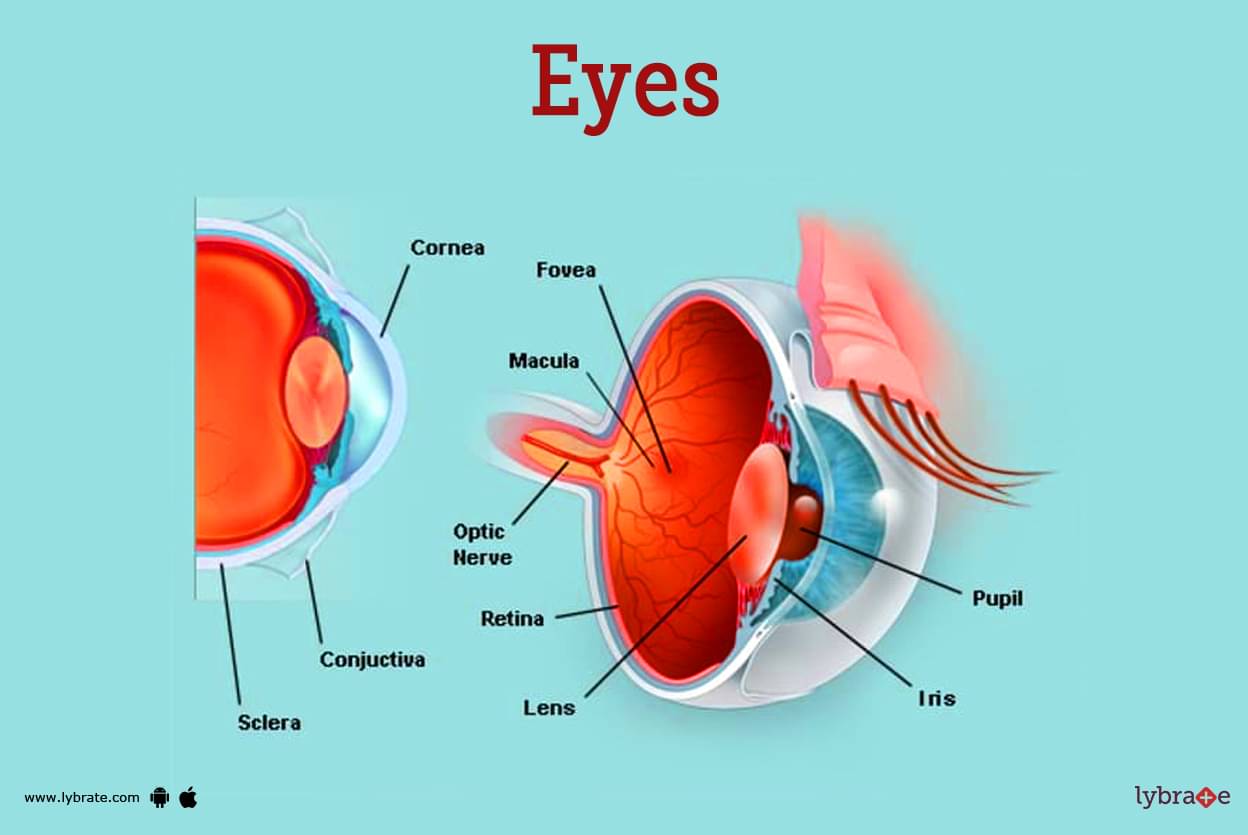
Our eyes are organs that allow us to see the world around us. They take in light from the environment around us and send visual information to our brain. In addition to seeing what's directly in front of you and to the sides, each of our eyes can also see around 200 degrees in all directions (referred to as peripheral vision). Eyes are naturally round and asymmetrical in shape. About one inch can be considered the diameter of the eye.
The following are the primary components of an eye:
- Iris: It is a tissue with a coloured appearance that can be found in the anterior region of the eye. Inside of this tissue can be found a number of pupils.
- Cornea: This translucent dome-shaped structure sits atop the iris and is of interest. The cornea and the iris help identify the anterior chamber from the posterior chamber that is between the iris and the lens.
- Pupil: The black, circular tissue that is found in the iris and is responsible for allowing light to enter the eye is called the pupil. The pupil acts as a conduit for communication between the two separate compartments.
- Sclera: The sclera is the white, outermost layer of the eyeball, and it can be found at the front of the eye. cornea: The cornea is the transparent layer that continues this layer.
- Conjunctiva: The conjunctiva is a much thinner layer of tissue than the cornea, but it covers the entire front region of the eye and lies directly beneath it.
- Lens: The lens is a peculiar biological structure that is found in the eye. It is a body that is biconvex, transparent, and 1 centimetre in diameter. It may be found in the space between the front and back of the eye, and has a thickness of around 4 millimetres.
- Ciliary Body: The ciliary body is the most enlarged portion of the vascular tunic. It can be found directly in the midst of the vascular tunic. It is continuous with the choroid that is behind it as well as the iris that is in front of it. It lies behind the corneoscleral junction, in front of the ora serrata of the retina, and it is a part of the sclera.
It has two separate chambers i.e, the smaller anterior chamber and the larger posterior chamber. The anterior chamber is the smaller of the two.
- Aqueous Humour: The aqueous humour contains a high concentration of amino acids, glucose, and ascorbic acid. It provides nutrition to the cornea and the lens, both of which are avascular otherwise.
- Choroid: The choroid is located in the back of the vascular coat of the eyeball, also known as the globe. The membranous layer that covers the inner surface of the sclera is brown, very thin, and densely vascularized. The ciliary body, which is located anteriorly, makes the connection to the iris, and the optic nerve, which is located posteriorly, pierces the ciliary body.
Function of Eye
The main functions of the eye are given below:-
- Capture light and convert it into electrical signals that can be interpreted by the brain. This process is known as vision.
- Adjust the amount of light that enters the eye through the pupil, which dilates or constricts in response to light levels. This helps to protect the retina from being damaged by too much light.
- Focus incoming light onto the retina at the back of the eye. The retina contains photoreceptors called rods and cones, which convert light into electrical signals that are sent to the brain via the optic nerve.
- Provide visual cues for depth perception and spatial awareness, which help us to navigate and interact with our environment.
- Help us to see color by separating light into its different wavelengths and detecting the relative intensity of each wavelength.
- Maintain eye health and protect the delicate internal structures of the eye from damage. The eye is equipped with various protective mechanisms, such as the eyelids, which help to keep dust and other foreign particles out of the eye, and the tear film, which helps to keep the surface of the eye moist and free of bacteria and other microorganisms.
- The primary purposes of the eye are to focus on light waves and to stimulate the photoreceptors that are located in the retina. This requires five fundamental processes, which are as follows:
- The passage of light waves through the transparent media that makes up the eyeball.
- The bending of light waves as they travel through various refractive media of varying densities.
- The ability of the lens to accommodate for changes in the distance in order to focus light waves
- The iris diaphragm is responsible for controlling the amount of light that comes into the eye through the pupil.
- The eyeballs coming closer together. When photoreceptors in the retina are stimulated, action potentials are produced. These action potentials are then transmitted to the visual cortex of the brain via the optic pathways. The visual cortex is where an image is formed. Visual impairment may be the end result if any one of these processes, or more than one of them, do not work as they should.
Disorders of eye
- Age-Related Macular Degeneration: Degeneration of the macula caused by ageing is referred to as age-related macular degeneration (AMD) and is a condition that can lead to vision loss.
- Amblyopia: Amblyopia is also referred to as 'lazy eye,' and it typically appears during childhood. As a result of this condition, one eye is able to see normally while the other eye, which is referred to as the 'lazy eye,' is unable to see normally. Because of this, the brain always gives preference to the more capable eye.
- Astigmatism: In the condition known as astigmatism, the eye is unable to properly focus on light because of irregularities in the cornea's natural curvature. This condition is treatable in a variety of ways, including eyeglasses, contact lenses, and even surgery.
- Black Eye: The term 'black eye' refers to the inflammation and discoloration that can appear around the eye as a result of a bruise. It's possible that the person has suffered some kind of facial injury that's causing this.
- Blepharitis: Blepharitis is an inflammation that can affect both the eyelids and the eyelashes, and it can be very uncomfortable. The area around the eye often feels gritty and itchy, which are both common symptoms of this condition.
- Cataract: This is a condition in which the internal lens of the eye becomes cloudy, causing a blurring of the patient's vision. Cataracts are very common.
- Chalazion: Chalazion is a condition in which the oil-producing glands of the eye become blocked due to an injury or other pathological action. This condition is known as chalazion. As a result, this leads to a blockage as well as swelling that forms a bump.
- Conjunctivitis: This condition is also commonly referred to as pinkeye. This condition is characterised by the presence of the infection, which results in inflammation of the conjunctiva. Allergies, viral or bacterial infections, or a combination of the two, are the most common causes of this disease.
- Corneal Abrasion: This condition occurs when an injury of some kind causes a scratch to appear on the cornea of the eye. As a result, the patient experiences the following symptoms: pain, sensitivity to light, and a feeling of grit in the eye.
- Diabetic Retinopathy: High blood sugar levels in the body can cause diabetic retinopathy, a condition in which the blood vessels in the eye become infected and damaged. Because of which the eye's vision is getting worse.
- Diplopia: The medical term for this condition is diplopia, which literally translates to 'double vision.' When someone has diplopia, their vision becomes double, which means that the object appears to be double due to a number of pathological as well as other serious conditions. Hence requires immediate medical attention.
- Dry Eye: Dry eye can also refer to the condition itself. Dry eye is typically the result of ageing, but it can also be brought on by certain medical conditions, such as lupus, scleroderma, and Sjogren syndrome.
- Glaucoma: This condition is caused by high pressure. Because it is so difficult to detect, it may go undiscovered for a number of years at a time. In situations like this, one's peripheral vision will be affected first, followed by their central vision.
- Hyperopia: This condition is also referred to as farsightedness. Because the lens is so small in this case, the eye is unable to see nearby objects clearly because either the light cannot properly penetrate the lens or the lens itself is too small. On the other hand, depending on the severity of the condition, one might experience difficulty with their distance vision.
- Hyphema: Hyphema is typically caused by trauma to the eye, which leads to bleeding into the front of the eye, specifically in the space between the iris and the cornea. This condition is known as hyphema.
- Keratitis: Keratitis is an inflammation of the cornea that occurs as a result of an infection. The majority of cases of this disease are brought on by the introduction of the pathogen through the cornea being scratched.
- Myopia: Nearsightedness is another name for this condition. On the other hand, the near vision might be affected or it might not. As a result of this condition, the lens has grown to an abnormally large size, which prevents light from being able to focus correctly on the retina.
- Optic Neuritis: Optic neuritis is characterised by inflammation of the optic nerve, which is typically brought on by an overactive immune system. This condition is responsible for both pain and a reduction in vision in one eye.
- Pterygium: This refers to the thickened mass that can be found in the anterior segment of the eyeball. Pterygium has the potential to cover up a portion of the cornea, leading to vision problems for the patient.
- Retinitis: It is a condition of the eye known as retinitis, which occurs when there is inflammation of the retina as a result of an infection, a long-term genetic disorder known as retinitis pigmentosa, or both.
- Retinal Detachment: When the retina detaches from its usual connection in the back of the eye, it causes a condition known as 'detachment of the retina.'. Retinal detachment is also known as detached retina. This condition is typically brought on by diabetes and trauma, and as a result, requires immediate medical attention in the form of surgical treatment.
- Scotoma: Scotoma is a condition of the eye that causes a blind spot in the vision.
- Strabismus: When the eyes are unable to point in the same direction, a person may be diagnosed with strabismus, a condition in which the brain gives preference to one eye over the other. It is possible for children to experience vision loss if they are affected by this condition. Amblyopia would be the consequence of this condition.
- Stye: The painful condition known as stye is caused by a bacterial infection that manifests as a red lump at the corner of the eyelid.
- Uveitis: In patients who have this condition, the coloured part of the eye will become inflamed as a result of an infection caused by a pathogen that is either bacterial or viral.
Test for eye
- Tonometry: Tonometry is a test that is performed in order to measure the pressure inside of the eye, which is referred to as intraocular pressure. Glaucoma can be evaluated with the help of this method, which is utilised by medical professionals.
- Slit Lamp Examination: slit lamp examination is a diagnostic tool that can identify a variety of eye conditions.During this examination, the doctor will examine the patient's eye using a microscope while shining a ray of light in a vertical direction around the eye.
- Fundoscopic Exam: An optometrist will perform a fundoscopic exam in order to look for any problems with the retina. This exam involves pupil dilation, which refers to the widening of the pupil using some special eye drops.
- Refraction: During the refraction portion of the exam, the optometrist will place a number of lenses in front of the patient's eye, one at a time, in order to determine the patient's prescription. Lenses are inserted into the eye to perform this procedure, which improves one's vision.
- Visual Acuity Test: The visual acuity test is a measurement that determines whether or not an individual's eyes are able to distinguish between the various contours and particulars of an object at a specified distance. It is recommended to carry out this test in a specific and consistent manner in order to spot any changes in the quality of the product.
- Fluorescein Angiography: it is a method of angiography in which a dye is injected into the vein of the eye and taken various pictures of the retina. A fluorescein angiography is the name given to this diagnostic technique.
- Regular Adult Eye Exam: A regular adult eye exam consists of a battery of tests similar to those described above, followed by an examination of how the eyes move together.
Treatments of Eye
- Contact Lenses And Glasses: The most common approach to treating common vision issues such as myopia, hypermetropia, and astigmatism is to use corrective lenses such as contact lenses and glasses.
- Lasik: LASIK stands for laser-assisted in-situ keratomileusis. During this procedure, your doctor will cut a small flap in your cornea and then use a laser to reshape your eye. The patient will undergo this procedure in order to correct vision issues such as excessive nearsightedness, farsightedness, and astigmatism.
- Photorefractive Keratectomy, Or PRK: To treat conditions such as nearsightedness, farsightedness, or astigmatism, this optometrist first rubs the top cells off of the cornea, and then uses a laser to remove the remaining cells. On the other hand, the surface cell was able to easily heal itself.
- Artificial Tears: Eye drops that contain artificial tears are designed to replicate the function of natural tears. It is used as a treatment for dryness as well as irritation of the eyes.
- Punctal Plugs: It is common practice to insert tiny plugs made of collagen or silicon into the ducts of the eye. These plugs are designed to keep tears in the eye for an extended period of time, thereby preventing dry eye syndrome and other eye complications.
- Cataract Surgery: Cataract surgery involves the removal of the eye's natural lens and its replacement with a lens that is manufactured artificially in order to clear up the cloudy vision that is caused by cataracts.
- Laser Photocoagulation: Laser photocoagulation is a treatment that is used by doctors to treat abnormal blood vessels by laying a laser on parts of the retina.The treatment for this issue involves the use of a laser. It is a common approach that is taken when treating circulation issues.
Medicines for Eyes
- Ceqa Eye Drops: Inflammation can be treated with ceqa eye drops, which belong to the cyclosporine drug class. Cyclosporine drugs have anti-inflammatory effects. Additionally effective in treating dry eyes.
- Restasis Eye Drops For Dry Eye: this is an example of the cyclosporine drug class, which is an anti-inflammatory action medication. These drops are used to treat dry eye syndrome. Additionally effective in treating dry eyes.
- Xiidra Eye Drops For Inflammation: These medications are included in the category of eye drops containing lifitegrast that are used to treat inflammation of the eye.
- Tyrvaya Spray For Basal Tears: These sprays contain varenicline, a medication that was just recently given the OK by the Food and Drug Administration and is used to stimulate the production of basal tears in the eye. These prescription drugs are to be taken twice daily.
- Aciclovir Eye Ointment: It uses antiviral eye ointment. Used to cure blurred vision of the eye.
- Acetazolamide For Glaucoma: It is a class of A carbonic anhydrase inhibitor. Used to treat glaucoma. Diamox are common brand names.
- Atropine Eye Drops: It is among An antimuscarinic class of drug.
- Azelastine Eye Drops For Allergies: It is commonly used in to make the pupil of your eye larger and relax the muscles in your eye
Table of content
Find Ophthalmologist near me
Ask a free question
Get FREE multiple opinions from Doctors


