

Brachiocephalic Vein (Human Anatomy): Image, Functions, Diseases and Treatments
Last Updated: Mar 18, 2023
Brachiocephalic Vein Image
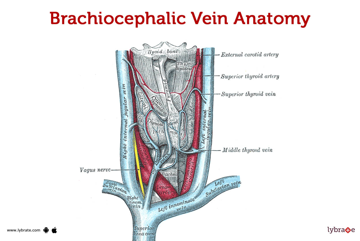
The blood in your head, neck, and arms is low in oxygen, but the brachiocephalic vein helps get it back to your heart. As a paired vein, it can be found on both the left and right sides of the human body (right and left). The brachiocephalic veins begin their journey at the point where the subclavian vein and the internal jugular vein meet.
Which veins form the brachiocephalic vein?
The brachiocephalic vein is formed when the internal jugular vein and the subclavian vein are connected to one another. The internal jugular vein is responsible for transporting blood from the head and neck, whereas the subclavian vein is in charge of transporting blood from the upper limbs. These two veins merge to produce the brachiocephalic vein, which then travels through the chest to the right atrium of the heart.
Where is the brachiocephalic vein located?
- The brachiocephalic vein can be found in the hollow of the thoracic cavity (chest).
- The internal jugular vein and the subclavian vein are both components of this vascular system.
- Blood from the head and neck is carried by the internal jugular vein, while blood from the upper limbs is carried by the subclavian vein.
- When these veins merge, they become the brachiocephalic vein and begin their course into the chest.
- It begins in the left atrium of the heart and makes its way through the thorax to the right atrium.
Which veins drain into the brachiocephalic vein?
The brachiocephalic vein is formed when the internal jugular vein and the subclavian vein are connected to one another. The internal jugular vein is responsible for collecting blood that has been drained from the head and neck, whereas the subclavian vein is responsible for collecting blood that has been drained from the upper limbs.
The brachiocephalic vein is formed when these veins converge; it then goes down the chest and into the right atrium of the heart. Oxygen-depleted blood is pumped back into circulation after travelling from the head, neck, and upper limbs via the brachiocephalic vein, a big vein.
.jpg)
How big is the brachiocephalic vein?
Your right and left brachiocephalic veins are very different from one another, which is a big difference. The length of the right brachiocephalic vein in your body is roughly 3 centimetres. Two aspirins side by side provide a useful mental image for this distance. The combined size of the two pills is over 3 centimetres.The brachiocephalic vein on your left side is approximately 6 centimetres longer than the one on your right.
What is the brachiocephalic vein made of?
The brachiocephalic vein has a layer of smooth muscle and connective tissue, with a layer of endothelial cells lining the inside.
Veins are supported and protected by smooth muscle and connective tissue, while blood flow is regulated by endothelial cells lining the interior of the veins. Through its course through the thorax, the brachiocephalic vein is protected by a layer of fat and connective tissue.
What anatomical variations affect the brachiocephalic vein?
Numerous anatomical variables have the potential to have an effect on the brachiocephalic vein.The occurrence of a second brachiocephalic vein is one example of this kind of diversity. An auxiliary brachiocephalic vein is present when a person has a second brachiocephalic vein. These supplementary veins are often smaller in size and can appear on either the left or right side of the body. Approximately 2% of the general population may be affected.
Persistent left superior vena cava is another kind that can influence brachiocephalic vein (LSVC). Blood from the head and upper limbs enters the LSVC on its way to the heart. The LSVC is usually replaced by the left brachiocephalic vein throughout development. About 0.5% of the population has this variant.
Another potential change to the brachiocephalic vein is the occurrence of a common brachiocephalic vein. This variant describes the situation in which the right and left brachiocephalic veins merge into a single vein. Roughly 1% of all people have this variant.
When taken as a whole, these anatomical variances can have a negative impact on the brachiocephalic vein's ability to function and structure, potentially resulting in issues like slowed blood flow or an increased risk of blood clots. However, in most cases, these variants are symptomless and have no noticeable impact on health.
Brachiocephalic Vein Functions
The blood that has been deprived of oxygen travels from the head, neck, and upper limbs back to the heart through a large vein known as the brachiocephalic vein. When the internal jugular vein and the subclavian vein come together, a new vein called the right atrial vein is produced. The brachiocephalic vein helps carry oxygen-depleted blood back to the heart, where it can be reoxygenated before being pumped to the rest of the body.
Brachiocephalic Vein Conditions and Disorders
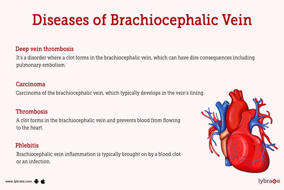
- Thrombosis: A clot forms in the brachiocephalic vein and prevents blood from flowing to the heart.
- Phlebitis: Brachiocephalic vein inflammation is typically brought on by a blood clot or an infection.
- Varicose veins: Venous varicosities of the brachiocephalic vein are varicose veins that produce pain and discomfort due to their enlargement and twisting.
- Venous insufficiency: Insufficient blood flow in the brachiocephalic vein, causing discomfort and swelling in the shoulder and neck.
- Deep vein thrombosis: It's a disorder where a clot forms in the brachiocephalic vein, which can have dire consequences including pulmonary embolism.
- Aneurysm: The brachiocephalic vein may become weakened or even burst if it develops a bulge in its wall.
- Arteriovenous malformation: A malformation in which an artery and vein form an improper connection within the brachiocephalic vein. Because of this, there is a higher chance of bleeding and irregular blood flow.
- Venous stenosis: It's a condition in which the brachiocephalic vein gets constricted and blood flow is impeded.When lymph fluid builds up abnormally in the brachiocephalic vein, this is called a lymphatic malformation. This might lead to discomfort and edema.
- Pulmonary embolism: Condition where a blood clot travels from the arm vein (brachiocephalic vein) to the lung.
- Superior vena cava syndrome: When the superior vena cava, the vein that carries blood from the head, neck, and upper body to the heart, becomes constricted or blocked, the outcome can be a reduction in blood flow as well as an increased chance of experiencing complications.
- Carcinoma: Carcinoma of the brachiocephalic vein, which typically develops in the vein's lining.
- Peripheral venous disease: Caused by obstruction in the brachiocephalic vein, this disorder causes blood to pool in the legs and arms.
- Venous thromboembolism: The formation of a blood clot in a vein, which can have life-threatening consequences including pulmonary embolism.
Brachiocephalic Vein tests
There are several tests that can be used to diagnose and evaluate the function of the brachiocephalic vein. These tests include:
- Ultrasound: In order to get a clear picture of the brachiocephalic vein, a non-invasive imaging technique called ultrasound is used. It can be utilised to detect any anomalies or blockages in the vein and evaluate its size, shape, and structure.
- CT scan: The brachiocephalic vein can be seen in great detail on a CT (computed tomography) scan, which uses X-rays to create an image. They are helpful for determining the health of the vein and determining whether any problems exist, such as blockages.
- Magnetic resonance imaging (MRI): MRI (magnetic resonance imaging) employs a powerful magnetic field and radio waves to obtain detailed images of the brachiocephalic vein. It can be used to examine the vein and determine its health and functionality.
- Venography: An X-ray image is taken before and after injecting a dye into the brachiocephalic vein for a diagnostic procedure called venography. It can evaluate the health of the vein and its ability to carry blood and discover any problems or blockages inside it.
- Angiography: Angiography is a diagnostic procedure in which a dye is injected into the brachiocephalic vein and its progress through the vein is photographed using X-rays. It can be used to evaluate the health of the vein and detect any problems, such as blockages.
- Phlebography: In phlebography, a diagnostic dye is injected into the brachiocephalic vein, and the vein's path is tracked using X-rays. It can be used to examine the vein and determine its health and functionality.
Brachiocephalic Vein Treatments
- Thrombolysis: Thrombolysis is a procedure wherein drugs are used to break up clots in the brachiocephalic vein. By increasing blood flow, it helps reduce the risk of cardiovascular problems like stroke and heart attacks.
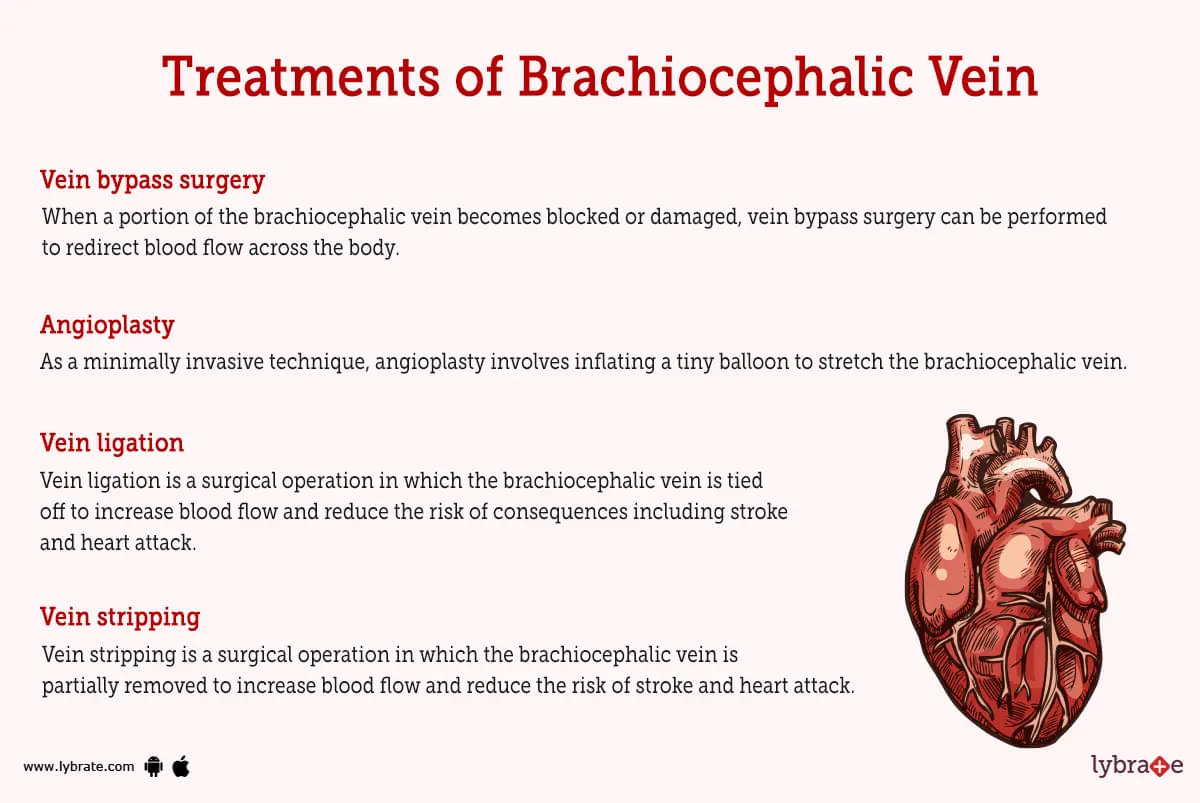
- angioplasty: As a minimally invasive technique, angioplasty involves inflating a tiny balloon to stretch the brachiocephalic vein. Its use can increase blood flow and reduce the risk of cardiovascular problems like stroke and heart attack.
- Stenting: Stenting is a minimally invasive treatment in which a tiny metal mesh tube is placed in the brachiocephalic vein to keep it open and enhance blood flow. Consequences like stroke and heart attacks can be avoided with its help.
- Vein ligation: Vein ligation is a surgical operation in which the brachiocephalic vein is tied off to increase blood flow and reduce the risk of consequences including stroke and heart attack.
- Vein stripping: Vein stripping is a surgical operation in which the brachiocephalic vein is partially removed to increase blood flow and reduce the risk of stroke and heart attack.
- Vein transposition: Vein transposition is a surgical operation used to enhance blood flow and prevent issues like stroke and heart attack by rerouting a portion of the brachiocephalic vein to another part of the body.
- Vena cava filter placement: Placing a metal filter in the vena cava (the big vein that conducts blood from the body to the heart) is a common operation for preventing blood clots from reaching the lungs and causing a heart attack or stroke. To reduce the danger of blood clots, it is used as an alternative to anticoagulant drugs.
- Vein thrombectomy: Blood clots in the brachiocephalic vein can be surgically removed with a treatment called a vein thrombectomy.
- Vein endarterectomy: Vein endarterectomy is a surgical operation used to remove plaque or other blockages from the brachiocephalic vein. As a result, it can be utilised to increase blood flow and avert potentially fatal conditions including stroke and heart attack.
- Vein bypass surgery: When a portion of the brachiocephalic vein becomes blocked or damaged, vein bypass surgery can be performed to redirect blood flow across the body. By increasing blood flow, it helps reduce the risk of cardiovascular problems like stroke and heart attacks.
How can I keep my brachiocephalic vein healthy?
- Sustaining a healthy lifestyle can help you keep your brachiocephalic vein in good condition. This includes avoiding the use of tobacco products, maintaining a healthy diet, and working out on a consistent basis. This includes not smoking, eating healthily, and keeping up with a regular exercise routine.
- Additionally, problems might be avoided by managing pre existing illnesses including hypertension and diabetes.
- Compression stockings, regular breaks to stretch and move about, and other similar measures can all aid in better blood circulation and the avoidance of blood clots. Talk to your doctor if you have any symptoms or concerns regarding your brachiocephalic vein so it can be diagnosed and treated correctly.
Brachiocephalic Vein Medicines
- Antihypertensive medications for high blood pressure in Brachiocephalic Vein: Medicines called antihypertensives are used to treat hypertension. As a result, blood vessel constriction and blood pressure drop, respectively. Lisinopril and amlodipine are examples of antihypertensive drugs that may be used to treat brachiocephalic vein hypertension.
- Diuretics for excess fluid in Brachiocephalic Vein: The term 'diuretic' refers to a class of drugs used to flush extra fluid from the system. They do what they're supposed to do by stimulating urination, which then removes the surplus fluid from the body. Hydrochlorothiazide and furosemide are two examples of diuretics that could be used to treat brachiocephalic vein edema.
- Anti-inflammatory medications for reducing inflammation in Brachiocephalic Vein: Swelling and inflammation can be diminished with the help of anti-inflammatory drugs. They reduce inflammation by blocking the body's generation of inflammatory substances. Naproxen and celecoxib are a couple of the aforementioned examples.
- Antidepressants for pain management in Brachiocephalic Vein: It has been suggested that antidepressants, which are typically used to treat depression and other mood disorders, may also be useful for managing chronic pain by blocking the brain's reception of pain signals. Medications like amitriptyline and venlafaxine are two such instances.
- Antidepressants for pain management in Brachiocephalic Vein: It has been suggested that antidepressants, which are typically used to treat depression and other mood disorders, may also be useful for managing chronic pain by blocking the brain's reception of pain signals. Medications like amitriptyline and venlafaxine are two such instances.
- Anticoagulants for preventing blood clots in Brachiocephalic Vein: Preventing the formation of blood clots is the primary purpose of anticoagulants. They achieve their effects by reducing the production of proteins critical to blood coagulation. Warfarin and heparin are two such examples.
- Beta blockers for lowering blood pressure in Brachiocephalic Vein: To reduce blood pressure, beta blockers work by preventing the hormones from doing their job and narrowing blood vessels. Medications like propranolol and metoprolol are good examples.
- Calcium channel blockers for lowering blood pressure in Brachiocephalic Vein: Medications known as calcium channel blockers are taken to reduce blood pressure by preventing calcium from entering vessel cells. Nifedipine and amlodipine are two such examples.
- Cholesterol-lowering medications for preventing blockages in Brachiocephalic Vein: Medication to reduce blood cholesterol levels is commonly utilised to forestall arterial plaque buildup. Staining and fibrates are two such examples.
Table of content


Find Vascular Surgeon near me

Ask a free question
Get FREE multiple opinions from Doctors