

Cartilage (Human Anatomy): Image, Functions, Diseases and Treatments
Last Updated: Mar 17, 2023
Cartilage Image
Your cartilage is the tough but flexible tissue that is found in many places in your body, including your joints. Elastic cartilage, fibrocartilage, and hyaline cartilage are the three primary forms of cartilage.
Elastic cartilage is very flexible and is found in your ear. Fibrocartilage is a bit tougher and is found in between your vertebrae in your spine and in your knee joint.
Protecting your joints and bones from damage is cartilage, a robust yet flexible connective tissue. Your entire body benefits from its ability to absorb shock.
By preventing the ends of your bones from grinding against one another, cartilage keeps your joints lubricated and functioning smoothly. In addition to being the primary structural tissue in some parts of your body, bone provides the basis for many of the organs and tissues you use every day.
Cartilage damage can develop suddenly, as in a sports injury or other trauma, or it can proceed gradually over time, leading to osteoarthritis. When cartilage is injured or destroyed, normal joint motion becomes difficult, if not impossible.
Where is cartilage located?
Cartilage pads the ends of bones almost everywhere they connect. And it's also at the ends of every bone in your body, where they come together to form joints.
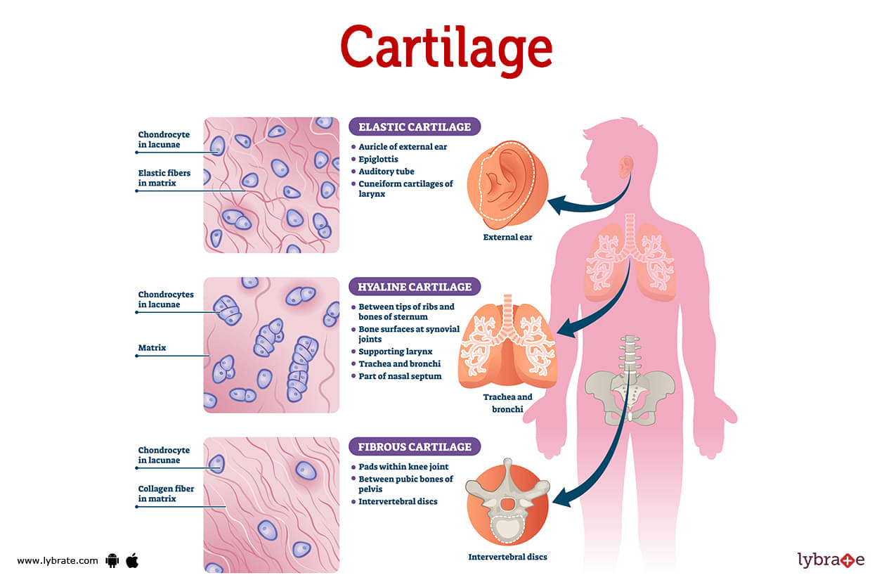
Types of cartilage
There are three types of cartilage in your body:
Hyaline cartilage
- Most of the cartilage in your body is hyaline cartilage. It covers the ends of the bones and acts as a lining for the joints. Bone ends are lined by hyaline cartilage, also known as articular cartilage.
- Because of its slippery and smooth nature, hyaline cartilage facilitates the free movement of bones within a joint. It can be bent without breaking, and it is sturdy enough to keep your joints from deforming.
- Hyaline cartilage is slick and smooth, allowing your bones to glide easily past each other in your joints. It's flexible but robust enough to keep your joints in place.
.jpg)
Fibrocartilage
- Fibrocartilage is exactly what the name implies: tough cartilage made of thick fibers. It is the most powerful and least flexible of the three. It may keep your body components in place and cushion falls.
- Locations of fibrocartilage in your body include: Your knee's meniscus. In the discs that sit between your vertebrae in your spine and also Muscles, tendons, and ligaments throughout your body are supported.
Elastic cartilage
- Your most flexible cartilage is elastic cartilage. It provides support to parts of your body that must bend and move in order to function. Even after a strong force, elastic cartilage can return to its original shape. Your ear is made of a flexible cartilage. It's pliable and may be bent and reshaped without breaking or injuring the user.
- Elastic cartilage is a type of cartilage that is flexible and elastic. It can be found in the following places on your body: your outer ears, the Eustachian tube, the larynx, and the epiglottis. This type of cartilage allows these parts of your body to move and bend.
Cartilage Functions
Cartilage serves as a barrier for your bones and joints. It protects the articular surfaces of your bones and provides padding for your joints. It is the function of cartilage to work:
- As a shock absorber for your bones and joints, cartilage helps make regular motions pain-free. making everyday activities more comfortable. It does an excellent job of dispersing impact energy and lowering the load on your skeleton. Imagine the difference between doing some jumping jacks in bare feet and wearing a pair of running shoes. Cartilage provides shock absorption in your joints and wraps around your bones like the padding in a good pair of sneakers.
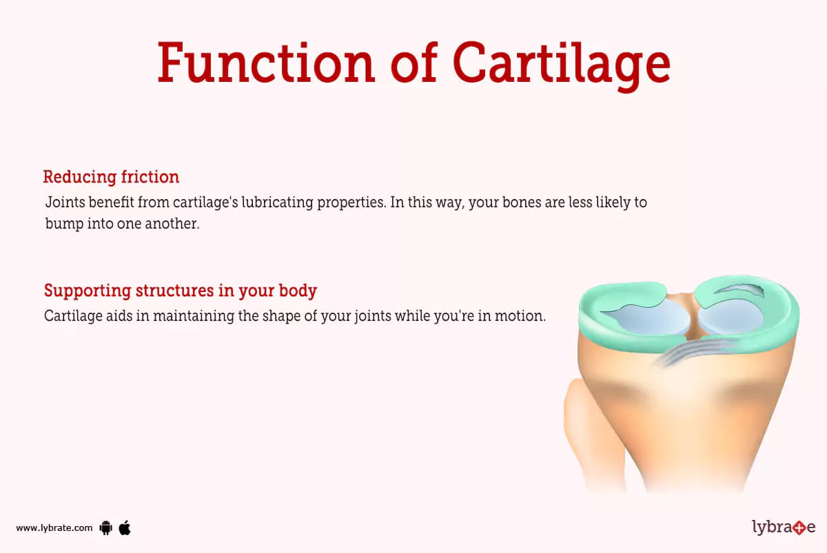
- Reducing friction: Joints benefit from cartilage's lubricating properties. In this way, your bones are less likely to bump into one another. Because of this, your joints can function normally, and their lifespan is extended.
- Supporting structures in your body: Cartilage aids in maintaining the shape of your joints while you're in motion. Furthermore, it joins various tissues and the skeletal framework together. Cartilage is linked to your muscles, tendons, and ligaments.
Cartilage is also the main tissue in some parts of your body including your Nose, Ears, Windpipe (your trachea).
Cartilage Conditions and Disorders
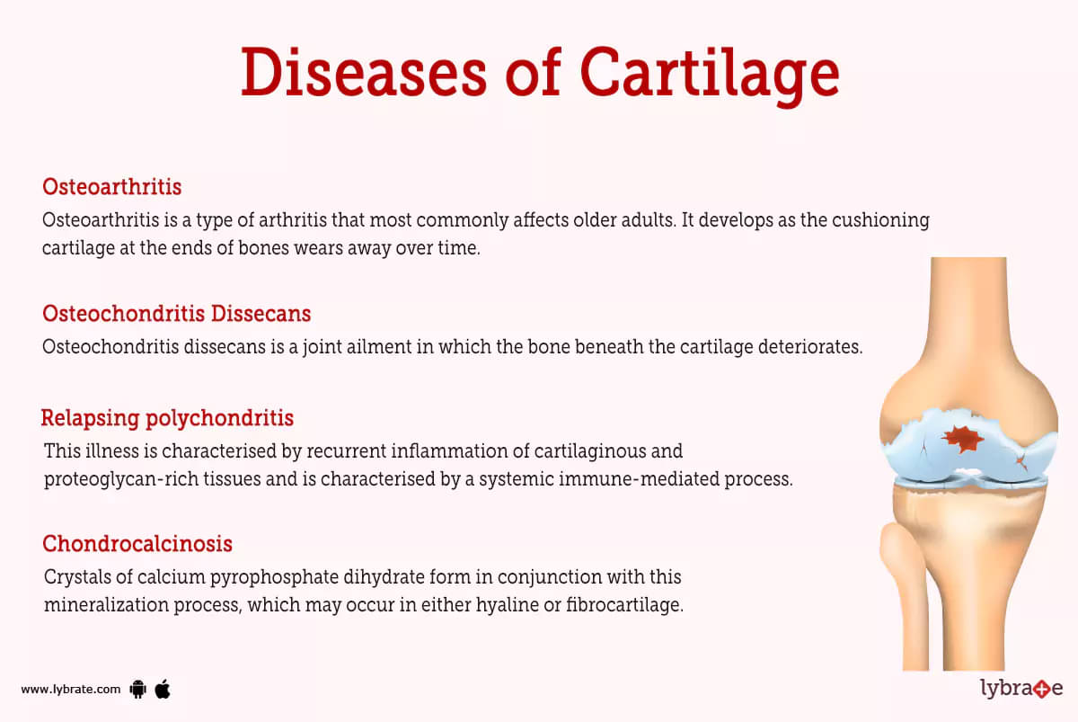
- Osteoarthritis: Osteoarthritis is a type of arthritis that most commonly affects older adults. It develops as the cushioning cartilage at the ends of bones wears away over time. The result might be aching, stiffness, and swelling in the joints. Everyday tasks, such as walking or climbing stairs, may become challenging for those suffering with osteoarthritis.
- Osteochondritis Dissecans: Osteochondritis dissecans is a joint ailment in which the bone beneath the cartilage deteriorates. This might result in joint discomfort, edema, and stiffness. In severe situations, the cartilage may become detached and float around in the joint, exacerbating the pain.
- Relapsing polychondritis: This illness is characterised by recurrent inflammation of cartilaginous and proteoglycan-rich tissues and is characterised by a systemic immune-mediated process. Because of the inflammation, the disease's structures gradually distort and become less functional.
- Chondrocalcinosis: Crystals of calcium pyrophosphate dihydrate form in conjunction with this mineralization process, which may occur in either hyaline or fibrocartilage. Chondrocalcinosis is most commonly found in the knee menisci.
- Chondrocalcinosis is distinguished by the following symptoms: This illness has symptoms with osteoarthritis and rheumatoid arthritis.
- Cartilage Tumours: Cartilage tumours Due to their resemblance to cartilage, head-and-neck cartilaginous tumours are rare yet difficult to diagnose. Head and neck benign cartilaginous tumours come in numerous varieties. Chondromyxoid fibroma, synovial chondromatosis, osteochondroma, and chondroma are examples.
- Inflammatory arthropathies: They cause aberrant biological processes to inflame joints and other tissues. Inflammatory arthropathy describes these conditions. Non-inflammatory arthritis is worn-out joints. .
- Synovial hyperplasia: An increase in the cellularity of the synovial membrane is what is meant by the term 'synovial hyperplasia.' This condition causes synovial thickness, which is a typical radiographic finding in the presence of synovitis.
- Torn meniscus: A healthy meniscus serves as a shock absorber and creates a smooth surface for your knee to slide on. A torn meniscus occurs when the meniscus is damaged. A tear in the meniscus makes it impossible for the knee to rotate, which leads to discomfort and a locking knee joint. Injuries to the meniscus are rather frequent, especially among sportsmen.
Cartilage Tests
- X-ray: Imaging the knee with this technique helps rule out more serious conditions like fractures or degenerative osteoarthritis as the source of your pain.
- MRI: The ability of an MRI to provide pictures of soft tissues aids your doctor in determining whether you have recent damage to the knee cartilage.
- Physical Examination: An ankle injury, sprain, or other condition may be diagnosed after a thorough medical examination.
Cartilage Treatments
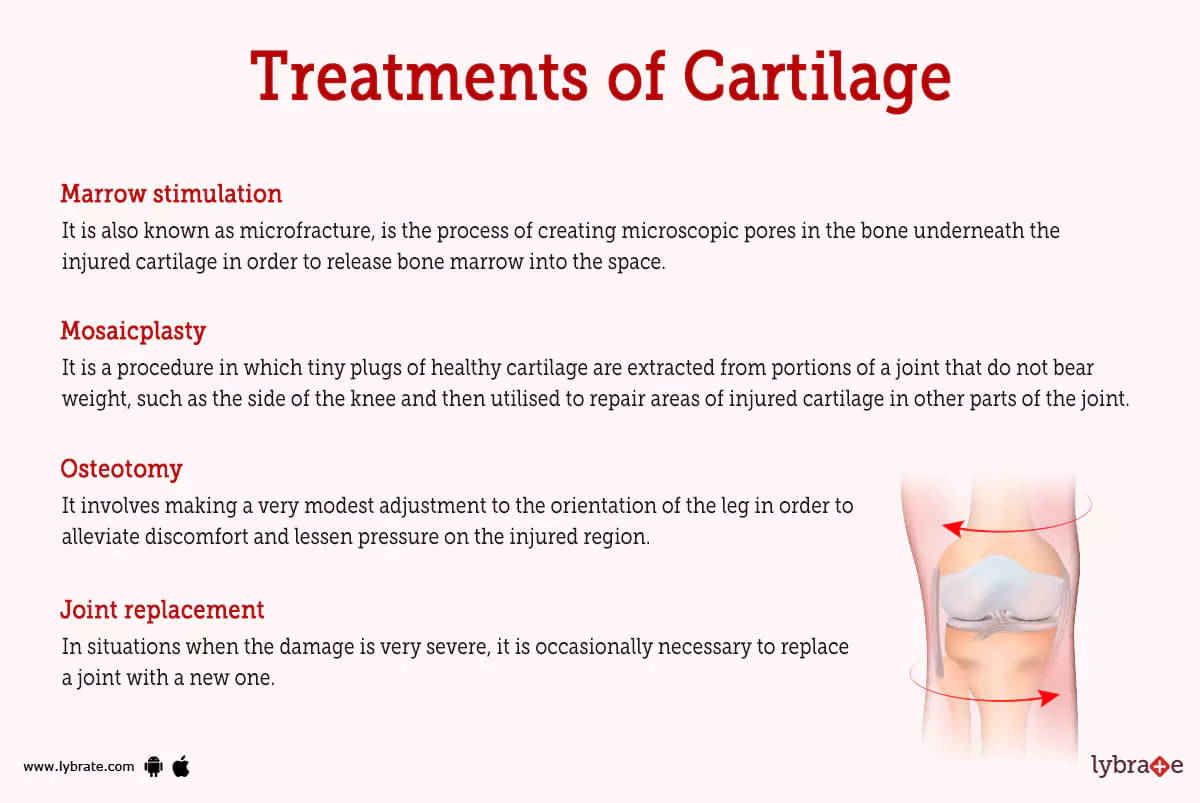
- Marrow stimulation: It is also known as microfracture, is the process of creating microscopic pores in the bone underneath the injured cartilage in order to release bone marrow into the space. These pores are produced in the bone during marrow stimulation. Following this, the cells in the bone marrow will start to encourage the formation of fresh cartilage.
- Mosaicplasty: It is a procedure in which tiny plugs of healthy cartilage are extracted from portions of a joint that do not bear weight, such as the side of the knee and then utilised to repair areas of injured cartilage in other parts of the joint.
- Osteotomy: It involves making a very modest adjustment to the orientation of the leg in order to alleviate discomfort and lessen pressure on the injured region. During this treatment, a wedge of bone is often added to or taken away from the shin or thigh bone. The bone is then secured with a plate while it heals.
- Joint replacement: In situations when the damage is very severe, it is occasionally necessary to replace a joint with a new one. This may include replacing the whole joint with an artificial joint, as is done when a patient's hip or knee is injured, as is the case with a hip or knee replacement.
- Allograft osteochondral transplantation: AOT is a surgery that is very similar to mosaicplasty. However, in allograft osteochondral transplantation (AOT), the replacement cartilage is obtained from a donor who has just gone away, and it is used to mend more extensive areas of damage.
- Autologous chondrocyte implantation: The initial step of the surgery known as autologous chondrocyte implantation, also known as ACI, is for the surgeon to take a small sample of cartilage cells from the joint that is being treated. After that, the cartilage cells are utilised to develop more cells in the laboratory, and the freshly generated cells are used to repair the cartilage that has been injured.
- Artificial scaffold: Synthetic support structures in order to repair the damaged cartilage, a specialised patch or gel may be put to the area. The artificial scaffold is a kind of therapy that may either be given in combination with marrow stimulation or on its own.
Cartilage Medicines
- Steroids for reducing inflammation of Cartilage: Steroids are a type of medication that can be helpful for cartilage. They work by helping to decrease inflammation and swelling. This can help to reduce pain and improve joint function. Steroids can be taken by mouth or injected into the joint.
- Analgesics for pain in Cartilage: analgesics are medications that help to relieve pain. For example, if you have a headache, you might take an aspirin. If you have a stomachache, you might take ibuprofen. and also naproxen sodium and paracetamol are useful as analgesics.
- Antibiotics for infection in Cartilage: Antibiotics can be used to treat a bacterial infection. Medicines are commonly used to treat infections such as cellulitis. Some of the medications include Vancomycin and cephalosporin (or cefepime if Pseudomonas is a concern), as well as azithromycin or doxycycline.
Table of content


Find Orthopedic Doctor near me

Ask a free question
Get FREE multiple opinions from Doctors