Collar Bone (Human Anatomy): Image, Function, Diseases, and Treatments
Last Updated: Feb 25, 2023
Collar Bone Image
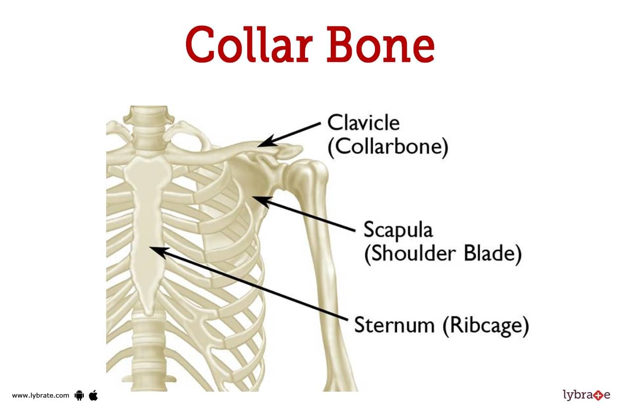
The clavicle, also known as the collarbone, is a long, slender bone with an S-shaped cross-section and a length of about 6 inches (15 cm). It serves as a link between the sternum and the scapula (breastbone).
One of the clavicles is on the left side, and the other is on the right side. The clavicle is the only other long bone in the body that is horizontal. It is part of the shoulder girdle and works in tandem with the should.er blade.
It is a bone that can be touched, and the placement of the bone is readily apparent in those who have less fat in this region because the bone generates a bulge in the skin. The bone is found in the lower part of the pelvic region. The bone was given the Latin name clavicula, which translates to 'small key,' since it turns along its axis like a key when the shoulder is abducted.'
The clavicle is the bone that gets fractured the most frequently. The bone is easily broken if there is a blow to the shoulder, such as when someone falls with their arms outstretched or takes a direct hit.
Collar Bone Function
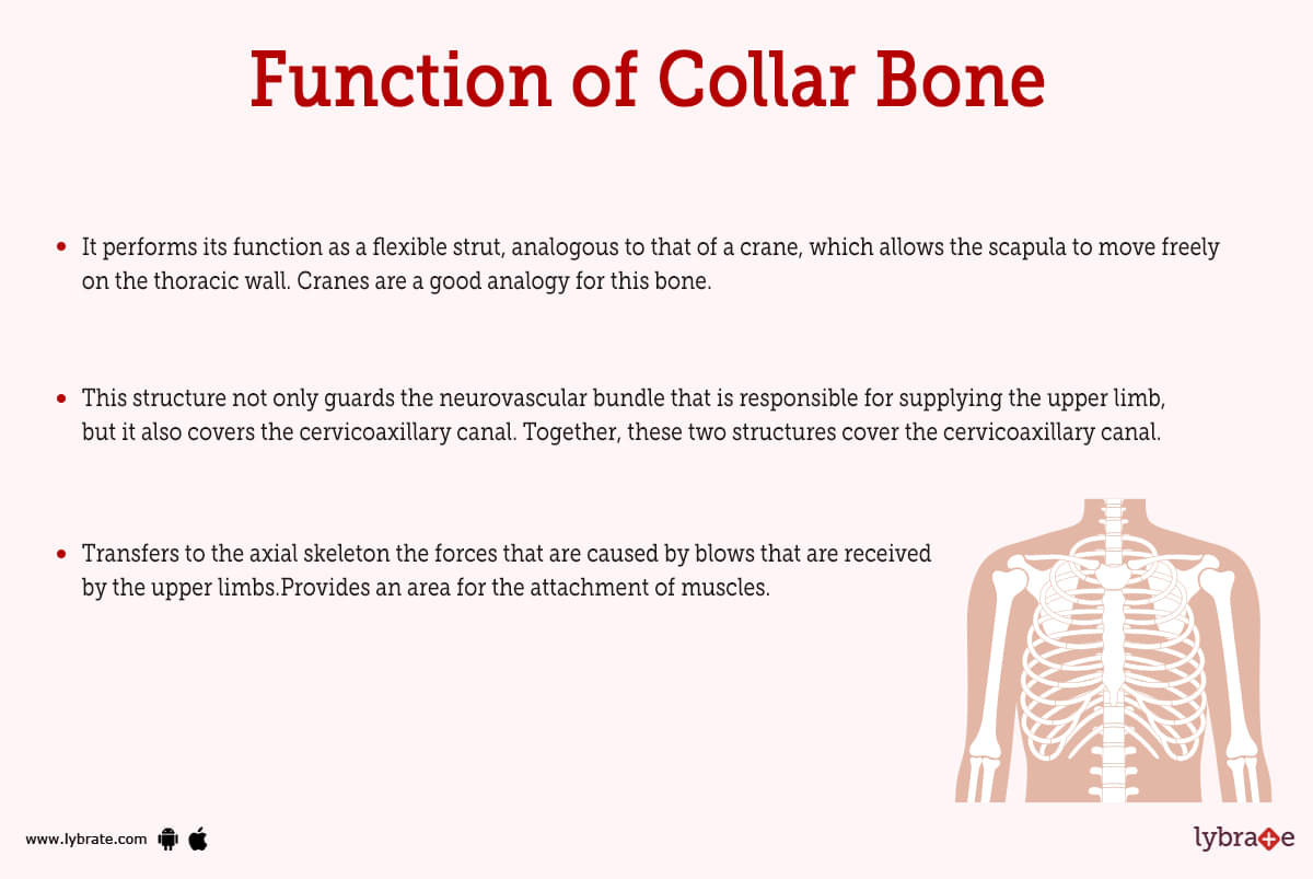
- This configuration maintains the upper limb away from the thorax in order to allow the arm to have the greatest possible range of motion, and it also fulfils the role of a hard support from which the scapula and the free limb are hanging.
- It performs its function as a flexible strut, analogous to that of a crane, which allows the scapula to move freely on the thoracic wall. Cranes are a good analogy for this bone.
- This structure not only guards the neurovascular bundle that is responsible for supplying the upper limb, but it also covers the cervicoaxillary canal. Together, these two structures cover the cervicoaxillary canal.
- Transfers to the axial skeleton the forces that are caused by blows that are received by the upper limbs.Provides an area for the attachment of muscles.
Collar bone Disorders
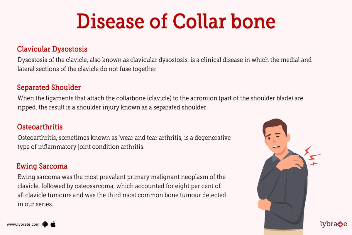
- Clavicular Dysostosis: Dysostosis of the clavicle, also known as clavicular dysostosis, is a clinical disease in which the medial and lateral sections of the clavicle do not fuse together. This is caused by the nonunion of two primary centres of ossification in the clavicle.
- Cleidocranial Dysostosis: Cleidocranial dysostosis is a clinical condition that is characterised by the absence of the clavicle, either partially or completely, in conjunction with abnormal ossification of the skull bones.
- Fracture Of Clavicle: Clavicle fractures are more prevalent than any other type of bone fracture. Fractures typically occur where the lateral and medial thirds of the bone meet. Due to direct impacts to the shoulder or indirect stresses, often caused by a severe blow to the hand or shoulder, the victim lands on the shoulder or the outstretched hand.
- Separated Shoulder: When the ligaments that attach the collarbone (clavicle) to the acromion (part of the shoulder blade) are ripped, the result is a shoulder injury known as a separated shoulder. The collarbone and the shoulder blade become farther distance from one another as a result of the rupture in the joint connecting the two bones. When someone gets a separated shoulder, the major ball-and-socket joint of the shoulder usually isn't harmed.
- Ewing Sarcoma: Ewing sarcoma was the most prevalent primary malignant neoplasm of the clavicle, followed by osteosarcoma, which accounted for eight per cent of all clavicle tumours and was the third most common bone tumour detected in our series. The fact that osteosarcoma is generally regarded to be the most prevalent primary malignant bone tumour in people makes this discovery even more remarkable.
- Dislocated Shoulder: When the humerus bone of the upper arm protrudes out of its normal articulation with the shoulder blade, this is known as a dislocated shoulder. In the case of partial dislocation of the humerus, some of the bone is still in its normal position within the socket. A dislocated humerus is one in which the bone has come completely loose from its articular socket.
- Osteoarthritis: Osteoarthritis, sometimes known as 'wear and tear arthritis, is a degenerative type of inflammatory joint condition arthritis. People who are 50 years of age or older are the ones who are most likely to be affected by it, but it can also strike people who are younger than that.The pain and stiffness that osteoarthritis causes gradually worsen over time as the disease progressively progress.
- Degradation Of Sternoclavicular Joint: When someone has osteoarthritis, the smooth articular cartilage that covers the sternoclavicular joint gradually wears away. This might result in a great amount of discomfort in the joint. As the cartilage deteriorates, it will eventually become ragged and rough, and the protective gap that is present between the bones will shrink. This can cause the bone to rub painfully against the other bone, and it can also develop a bony prominence surrounding the joint.
- Bursae Of Clavicle: Bursitis is the medical term for an inflammation of the bursae (also known as a bursal sac). Bursae are small sacs that are filled with fluid and are located close to joints. They prevent the tendons, muscles, and bones in the joint from rubbing against one another by acting as a cushion between the moving parts of the joint. If a bursa in the shoulder becomes irritated, it will become inflamed, and it will also grow in size. Because of this, the shoulder has less space for the muscles and tendons to move around, which can cause discomfort as well as a reduction in the range of motion in the shoulder.
- Distal Clavicular Osteolysis: Pain in the AC joint is often the result of an overuse injury known as distal clavicular osteolysis (DCO). This injury is most frequently seen in athletes and weightlifters. This is an uncommon injury that can be successfully treated using a variety of approaches.
- Osteomyelitis: Osteomyelitis of the clavicle is an extremely uncommon condition. Infection occurs from hematogenous spread or trauma. When it comes to adults, infection is typically secondary and the result of an exogenous cause such as an open fracture, surgery (iatrogenic), or the spread of infection from infected local tissue.
- Thoracic Outlet Syndrome: This happens when the collarbone moves out of its normal position and presses on the blood vessels and nerves that are located in the space between the bone and the highest rib. This can be very painful.
The following are some of the potential causes of thoracic outlet syndrome:
Obesity, injury, poor stance, muscular weakness in the shoulders, impairment caused by frequent heavy lifting or strain congenital condition.
- Malunion: Malunion is the term used by medical professionals to describe the condition that occurs when bone fragments migrate improperly and then repair in the wrong location. It's possible you'll need surgery.
- Wound Problem: Problems with the wound The area of your skin where the surgeon made the incision may have issues healing, become infected, or haemorrhage.
- Nerve Injury: During surgery, patients frequently sustain damage to the skin nerves that are responsible for providing sensation just below the collarbone. However, major nerve damage is extremely rare. Patients who have undergone surgery for clavicle fractures frequently report feeling a patch of numbness or tingling directly under the location of their incision. It is possible that over time it will diminish in size and become less apparent; however, it is likely to continue existing.
Collar Bone Test
- Physical Exam: Exams of the patient's physical condition: A doctor can typically diagnose bursitis by examining the patient's range of motion in the shoulder joint, the strength of the patient's muscles, and the location of the pain.
- Radiography: An X-ray does not look at the bursa itself, but it can be used to rule out bone damage or arthritis as possible causes of shoulder pain. Radiography is a form of imaging that uses electromagnetic radiation. This is something that should be taken into consideration when selecting the appropriate form of treatment.
- MRI Scans: An MRI scan can show whether or not there is inflammation in the bursa, as well as whether or not there is damage to the bone and the surrounding tissue. MRI scans are typically unnecessary during the diagnostic process.
- Bone Marrow And Fluid Aspiration And Testing: Testing the Fluid Fluid can be drawn from the bursa and tested to determine whether or not an infection is present. This process is referred to by its proper term, aspiration.
- DEXA SCAN: A dual-energy x-ray absorptiometry (DEXA) scan is the method that is used most frequently and provides the most reliable results. Low-dose x-rays are used in DEXA imaging. It is one of the gold standards for checking the fractures and symptoms of arthritis.
- Bone Mineral Density (BMD): With the help of this test a health care practitioner can diagnose the composition of bone which can help in diagnosing the problems related to the bone.
.jpg)
Collar Bone Treatment
- Physical Therapy: A popular form of treatment is called physical therapy. The muscles that surround the collarbone will most likely be the primary focus of this workout. However, if the condition is severe enough, surgery can be necessary.
- Physiotherapy: When the clavicle is damaged due to injury, illness, or disability, physiotherapy can assist restore motion and function to the shoulder joint. More than that, it can help lessen the chances of you getting sick or hurt in the future. It calls for a comprehensive approach that maximally involves the patient in shaping his or her own treatment.
- Steroid Injections: Ultrasound-guided steroid injection into the acromioclavicular joint requires only a tiny amount of steroid but has been shown to significantly reduce swelling, irritation, and pain. Low doses of steroids are used in injectable forms. This injection was met with universally favourable responses from patients.
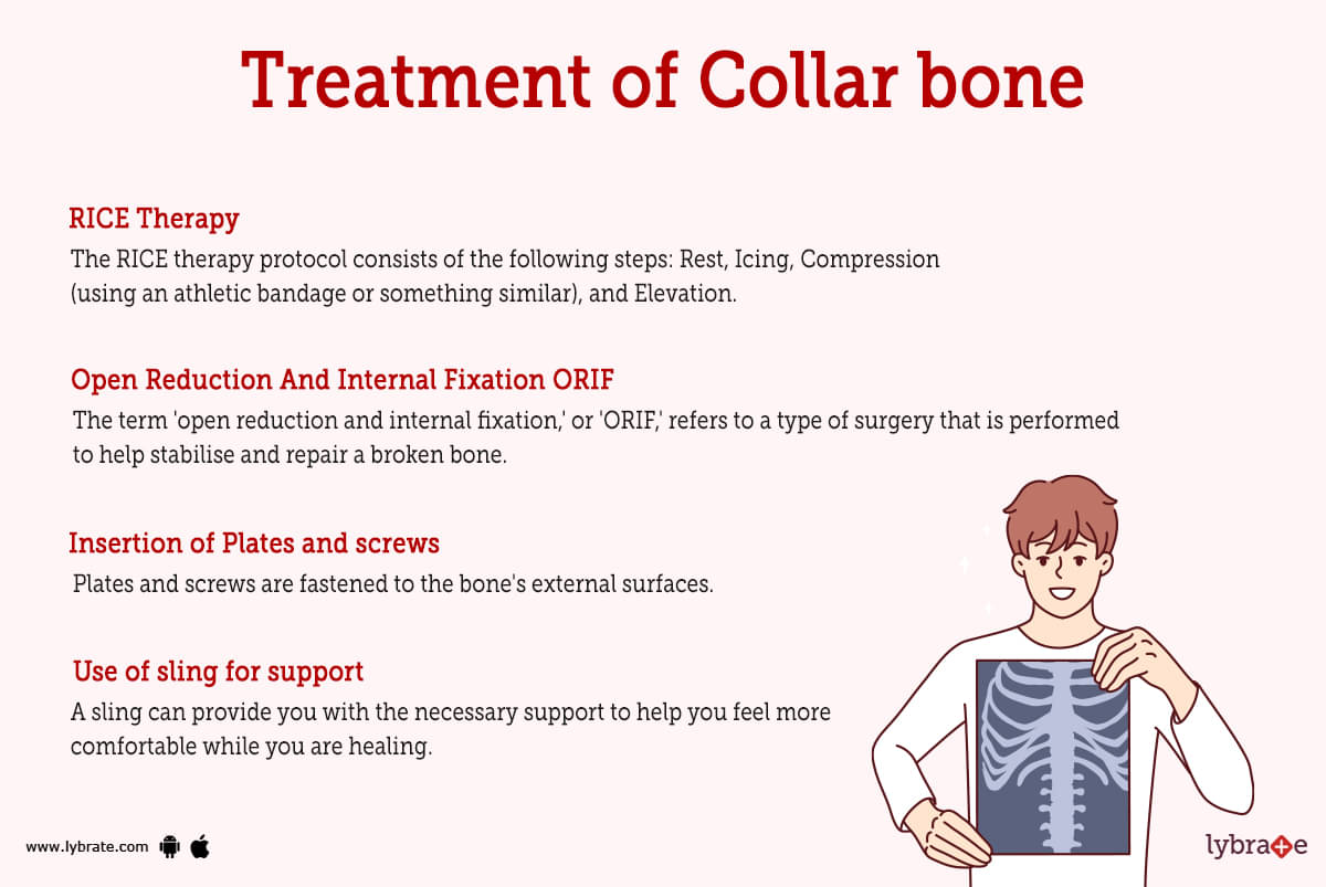
- RICE Therapy: The RICE therapy protocol consists of the following steps: Rest, Icing, Compression (using an athletic bandage or something similar), and Elevation. An injury to the foot can typically be treated using the RICE method, which consists of applying ice, resting the injured foot, applying compression, and elevating it.
- Open Reduction And Internal Fixation ORIF: The term 'open reduction and internal fixation,' or 'ORIF,' refers to a type of surgery that is performed to help stabilise and repair a broken bone. It's possible that you'll require this operation to treat the fracture in your collarbone (clavicle).
- Use of sling for support: A sling can provide you with the necessary support to help you feel more comfortable while you are healing. Additionally, it can stop the fractured pieces of bone from migrating around in the body.
- Insertion of Plates and screws: Plates and screws are fastened to the bone's external surfaces. In most cases, the hardware is not removed once the bone has healed, unless it is causing the patient distress (this usually happens a year or more after the surgery). Insertion Of Screws Or Pins: screws or pins that are driven through the bone. After the fracture has sufficiently healed, they are often removed.
- Surgery Of Clavicle Bone: In the vast majority of cases, an incision is created down the length of the collar bone, the fracture is exposed, the pieces are reattached to one another and fastened with the help of a combination of metal screws and a metal plate, and then the collar bone is reopened. The incision is then stitched shut using sutures that dissolve on their own, and the patient is then slung. The majority of patients are able to go home on the same day as their procedures.
Collar Bone Medicine
- Treatment For Fungal Infection: a topical antifungal medicine or by taking an oral antifungal drug. Luliconazole, itraconazole, clotrimazole, fluconazole, etc. are just a few examples used to treat fungal infection near the clavicle.
- Nonsteroidal anti-inflammatory drugs for pain in the collar bone: Nonsteroidal anti-inflammatory drugs (NSAIDs) are commonly used to treat clavicle pain, in addition to aches and pains in other parts of the body. Normal drugs such as ibuprofen, aspirin, and naproxen sodium fall within this category. Other NSAIDs include indomethacin, ketorolac, diclofenac, meloxicam, and celecoxib.
- Injections of Platelet-Rich Plasma (PRP) are used to treat joint pain: typically in the shoulder . PRP contains a number of growth factors. Platelet-rich plasma, or PRP, is another name for this plasma. This has many benefits, including reducing inflammation and encouraging the body's natural ability to heal injured tissue.
- Supplements for collar bone growth: Supplements designed to stimulate clavicle bone development through nutrition Nutritional supplements like glucosamine and chondroitin are commonly prescribed by doctors to ease their patients' pain and hasten the recovery of their joints.
- Vitamins and calcium supplements for collar bone fracture: Depending on the patient's age and the availability or deficiency of the necessary elements for normal bone formation and metabolism, vitamin D and calcium supplements may be administered.
- Neuropathic Pain Treatment For Osteomyelitis Of Collar Bone: Pregabalin, an anticonvulsant used to treat neuropathic pain and fibromyalgia, has been demonstrated to alleviate clavicle pain that radiates from the extremities. It is possible to use it as a standalone therapy or in combination with other seizure medications in order to treat partial-onset seizures.
- Medicines For Tumours: helpful in relieving clavicle pain caused by tumours Medications such as Allopurinol, which blocks xanthine oxidase, Febuxostat, which blocks xanthine oxidase, Probenecid, which blocks tubular resorption of uric acid in PCT Pegloticase, and Rasburicase, a recombinant uricase that converts uric acid to a water-soluble form, are all useful in the treatment of Gout and Tumours.
Frequently Asked Questions (FAQs)
What causes pain in the collarbone?
How serious is a collarbone fracture?
What causes cleidocranial dysplasia?
What is Cleidocranial Dysostosis?
What is the fastest way to heal a collarbone?
Can a collarbone heal without surgery?
How do you treat a collarbone at home?
What can cause collarbone pain without injury?
Can collarbone pain be heart-related?


Table of content


Find Orthopedic Doctor near me

Ask a free question
Get FREE multiple opinions from Doctors