Esophagus (Human Anatomy): Image, Function, Conditions, and More
Last Updated: Apr 08, 2023
Esophagus Image
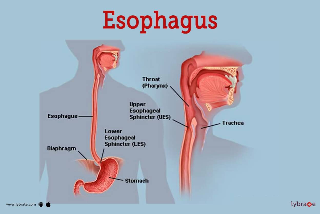
The Esophagus is a muscular tube that measures approximately 25 centimeters in length and connects the pharynx to the stomach. The Esophagus is maintained in a collapsed state between the trachea and the vertebral column in an anterior-posterior direction. It is only when a bolus of food goes through it that its size increases.
The Esophagus develops from a continuation of the pharynx at the lower border of the cricoid cartilage, which is located opposite the lower border of the Co vertebra.
It then travels downward in front of the vertebral column behind the trachea, through the superior and posterior medias tina of the thorax, through the esophageal opening of the diaphragm, and finally terminates at the cardiac orifice of the stomach in the abdomen about 2.5 centimeters to the left of the median plane. There are three distinct sections that make up the Esophagus: the cervical, the thoracic, and the abdominal sections.
- Upper esophageal sphincter: The upper esophageal sphincter will open reflexively upon swallowing for 0.2-0.3 seconds after the beginning of a swallow and will remain open for 0.5-1 seconds after the swallow has been completed. This makes it possible for the 'bolus' to enter the body of the Esophagus. Because it is normally disconnected from the pharynx, it prevents the swallowing of air while the body is respiration.
- Lower esophageal sphincter: The sphincter relaxes reflexively between 1.5 and 2.5 seconds after being stimulated by swallowing or the distension of the Esophagus caused by eating. This controls how quickly food travels from the throat to the stomach after being swallowed. The sphincter contracts and may go through a strong and prolonged contraction when peristaltic contractions reach this region. This prevents the regurgitation of food, gastric juice, and air. The LES muscles are not under voluntary control.
The development of peristalsis can be caused by swallowing as well as by local stimulation of the Esophagus at any level.
Functions of Esophagus
Its main functions include:
- Transporting food: The esophagus moves food from the mouth to the stomach through a process called peristalsis, where coordinated contractions of the esophageal muscles push the food downward.
- Protection against reflux: The lower esophageal sphincter is a ring of muscle located at the bottom of the esophagus that helps to prevent stomach acid and food from flowing back into the esophagus, a condition called gastroesophageal reflux disease (GERD).
- Coordination with other digestive organs: The esophagus works in coordination with the other organs of the digestive system, including the mouth, stomach, and small intestine, to help break down and absorb food.
- Help swallowing: Muscles in the esophagus help in moving the food bolus down to stomach
Esophagus Conditions
- Heartburn: Heartburn is caused by a weakened lower esophageal sphincter (LES), which allows stomach acid to reflux (back up) into the food pipe. This is the most common cause of heartburn. Acid reflux can show its face in a number of different ways, including heartburn, a persistent cough, a hoarse voice, or even in the absence of any symptoms at all.
- Oesophagitis: Inflammation of the Esophagus, also known as esophagitis. Esophagitis can be brought on by irritation (from things like reflux or radiation treatment), or it can be brought on by an infection.
- Barrett's Esophagus: The development of Barrett's Esophagus takes place when the lower portion of the Esophagus goes through structural alterations as a result of persistent irritation from stomach acid reflux. Very infrequently, Barrett's Esophagus can lead to the development of esophageal cancer.
- Esophageal ulcer: An ulcer in the lining of the Esophagus is referred to as an esophageal ulcer. In many instances, the problem is due to prolonged reflux.
- Esophageal stricture: Esophageal constriction is sometimes referred to as esophageal narrowing. In most cases, esophageal strictures are brought on by continuous discomfort brought on by reflux.
- Esophageal cancer: Cancer of the Esophagus is not very common but poses a significant threat to patients. A higher esophageal cancer risk is associated with smoking cigarettes, drinking excessive amounts of alcohol, and having acid reflux over an extended period of time.
- Mallory-Weiss tear: The Mallory-Weiss rip is a condition that develops when the lining of the Esophagus is torn due to vigorous vomiting or retching. The symptoms of nausea and vomiting can be brought on by a rupture in the Esophagus, which allows blood to stream into the stomach.Esophageal varices are swollen veins in the Esophagus, and cirrhosis can be the cause of this condition. Varices are a type of vein that is prone to rupture, which can result in significant internal bleeding.
- Esophageal ring (Schatzki's ring): An esophageal ring, also known as Schatzki's ring, is a common tissue aggregation that takes the form of a ring and is found around the base of the Esophagus. It is completely harmless. It's possible that having Schatzki's rings could make swallowing difficult, but that's about the worst thing that could happen.
- Esophageal web: An esophageal web is a term that refers to the aggregation of tissue that normally takes place in the upper Esophagus and is quite similar to an esophageal ring. Esophageal webs, much like esophageal rings, frequently go unnoticed by patients.
- Plummer-Vinson syndrome: Symptoms of Plummer-Vinson syndrome include anaemia, esophageal webs, and problems swallowing. Esophageal web dilatation and iron replenishment are also treatments that can be utilised.Swallowing can be made harder by a condition known as esophageal stricture, which refers to a narrowing of the Esophagus.
- Deglutition reflex: In its absence, it may result in the aspiration of food into the larynx or the regurgitation of food into the nose.
- Aerophagia: Due to the low resting tone of the upper esophageal sphincter that is characteristic of anxious people, it is impossible to avoid swallowing air while eating or drinking.
- Achalasia Cardia: This condition is distinguished by the inability of the lower esophageal sphincter to completely relax during swallowing as a result of the destruction of the local nerve plexuses. As a consequence of this, the number of neurons in the lower Esophagus that contain nitric oxide and VIP decreases, and the sphincter becomes more sensitive to circulating gastrin. The accumulation of food in the Esophagus, which is caused by a constriction of the sphincter, results in the proximal Esophagus expanding.
- Gastroesophageal reflux disease (GERD): When the lower esophageal sphincter is weak, stomach acid flows back up into the Esophagus, causing symptoms including heartburn and oesophagitis (gastroesophageal reflux disease-GERD). Ulceration and stricture from scarring can result.
- Dysphagia: It indicates that you have trouble swallowing. Disorders that affect any of the stages of the swallowing process can cause this condition.
Esophagus Tests
- Upper endoscopy, EGD (esophagogastroduodenoscopy): In upper endoscopy, also called EGD (esophagogastroduodenoscopy), a patient has an endoscope—a flexible tube with a camera attached to one end—inserted into his or her mouth. The endoscope provides simultaneous visualisation of the Esophagus, stomach, and duodenum (small intestine).
- Esophageal pH monitoring: Esophageal pH is monitored by introducing a probe into the Esophagus and measuring the acidity of the contents there (pH). pH testing can aid in the diagnosis of GERD and can be used to assess the effectiveness of treatment.
- Barium swallow: In order to do an X-ray examination of the Esophagus and stomach, a barium swallow requires the patient to drink a barium solution. A barium swallow is frequently used to diagnose the root of swallowing problems.
- CT Scan: Using computers and spinning X-ray machines, a CT scan produces cross-sectional images of the body. These scans provide greater detail than regular X-rays. They can show the skeleton, arteries, and other soft tissues in different parts of the body.
- Esophageal manometry: The pressure within the Esophagus is measured using esophageal manometry. It may assess the activity of the stripping muscle waves in the esophagus's major section as well as the muscle valve at the end.
- Esophageal impedance: The existence and severity of gastroesophageal reflux can be determined using esophageal impedance testing, which is performed in the Esophagus (the tube linking the neck and the stomach).
Esophagus Treatments
- Esophagectomy: Surgical removal of the esophagus, usually for esophageal cancer.Esophageal dilation: A balloon is passed down the esophagus and inflated to dilate a stricture, web, or ring that interferes with swallowing.
- Esophageal variceal banding: During endoscopy, rubber band-like devices can be wrapped around esophageal varices. Banding causes varices to clot, reducing their chance of bleeding.
- Biopsy: Often done through an endoscope, a small piece of the esophagus is taken to be evaluated under a microscope.
- Confocal laser endomicroscopy: A new procedure that takes the microscope inside a patient, which may replace the need for many biopsies.
Esophagus Medicines
- Histamine (H2) blockers for hyperacidity: The histamine enhances secretions of the stomach acid; blocking histamine decreases secretions of acid production and GERD symptoms etc.
- Proton Pump Inhibitors for GERD: These are responsible for the direct inhibition of the acid pumps in the stomach. It is taken daily to make it effective against stomach acidity problems. Though, some side-effects are reported by taking proton pump inhibitors for several months.
- Chloride Channel Activator for GERD: Lubiprostone is part of the clinical category known as chloride channel activators. This is accomplished by boosting the amount of fluid that is already present in your intestines, which, in turn, makes it easier for faeces to go through your digestive tract.
- Prebiotics for hyperacidity: In the therapeutic treatment, they are used to boost the activity of beneficial bacteria in the Esophagus, which allows for a more stable pH level in the Esophagus to be maintained as required.
- Antibiotics for oesophagitis: Antibiotics, in combination with other treatments, may be used to treat an infection caused by H. pylori. During treatment, the antibiotics are given to the stomach in an effort to heal the damage caused by the infection.
- Antiparasitic drugs: In the treatment of parasites, some examples include metronidazole, praziquantel, and albendazole. In the treatment of bacterial infections, some examples include azithromycin, ciprofloxacin, and tetracycline.
- Antiviral Medications: A number of antiviral drugs, such as entecavir, tenofovir , lamivudine , adefovir , and telbivudine, can aid in the fight against the virus and lessen its ability to harm your Esophagus.
- Chemotherapeutic Drugs: Chemotherapy and radiation are effective treatments for Esophagus cancer, despite the disease's untreatable nature. In extreme cases, the Esophagus may be removed surgically or replaced with a donor organ.
Frequently Asked Questions (FAQs)
What are signs of esophagus issues?
What causes esophagus damage?
Can esophagus be cured?
What are the 3 parts of the esophagus?
What does a damaged esophagus feel like?
What is the most common problem with the esophagus?
What foods heal the esophagus?
What is the fastest way to cure esophagitis?
What will heal my esophagus?
Table of content
Find Gastroenterologist near me
Ask a free question
Get FREE multiple opinions from Doctors


