

Iris (Human Anatomy): Image, Functions, Diseases and Treatments
Last Updated: Mar 18, 2023
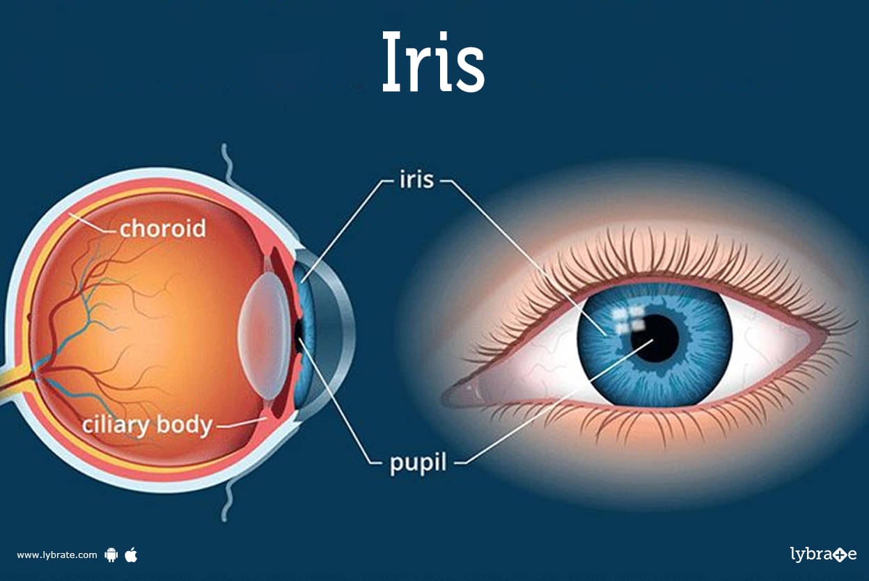
Iris Image
The color of your iris is unique, just like your fingerprint. Talk to your healthcare provider if you ever notice any changes in your vision or if you’re suddenly sensitive to changes in lighting.
The iris is the colored part of your eye. Muscles in your iris control your pupil — the small black opening that lets light into your eye.
The color of your iris is like your fingerprint. It’s unique to you, and nobody else in the world has the exact same colored eye.
Where is the iris in the eye?
The iris surrounds the pupil at the center of your eye.
Your eyeball has several layers that sit on top of each other, like an onion. The iris is one layer from the outside — under the cornea on top of the lens.
What does it look like?
The iris is the part of your eye that’s colored. It’s flat and round.
Your eye color depends on how much melanin (a naturally occurring pigment) your body makes and certain genes. The genes that determine your eye color are passed down through your parents.
What is the iris made of?
The iris is made of muscles and nerves. The nerves and muscles in your iris work on their own without you thinking about them (parasympathetically) to control the size of your pupil.
Your iris is filled with a fluid called aqueous humor. Your eye constantly produces and drains aqueous humor to maintain its shape, size and pressure.
.jpg)
Iris Functions
Muscles in your iris control your pupil. When your pupil is wider (dilated), more light gets into your eye. When it’s narrower (contracted) less light gets in.
As your iris squeezes or releases your pupil the amount of light reaching the rest of your eye changes. This constant change in size helps you see in different lighting. You’ve experienced this if you’ve stepped outside on a bright day or come inside after some time in the sun. The time it takes your eyes to adjust to the light is your irises adjusting your pupil to help you see.
How does the iris help the eye function?
The iris controlling your pupil helps your eyes see clearly. The iris is constantly changing how dilated your pupil is without you controlling it. This is called the pupillary light reflex.
Some people are born without an iris in one or both of their eyes — a genetic condition called aniridia. Without an iris, your eye would still function, but your vision would be blurry.
Iris Conditions and Disorders
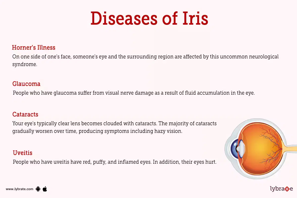
- Horner's Illness: On one side of one's face, someone's eye and the surrounding region are affected by this uncommon neurological syndrome. There are several potential reasons, ranging from carotid artery dissection to apical lung tumour, and it is a symptom of underlying nerve injury.
- Glaucoma: People who have glaucoma suffer from visual nerve damage as a result of fluid accumulation in the eye. This eye pressure may permanently impair eyesight if it is not addressed. The second most common cause of blindness worldwide is glaucoma.
- Cataracts: Your eye's typically clear lens becomes clouded with cataracts. The majority of cataracts gradually worsen over time, producing symptoms including hazy vision.
- Uveitis: People who have uveitis have red, puffy, and inflamed eyes. In addition, their eyes hurt. Uveitis is more likely to develop in certain circumstances, although it also often happens for unknown reasons. Treatments may help you regain your eyesight and stop it from happening again. After therapy, certain uveitis forms might recur. Uveitis left untreated might result in blindness.
- Chronic unilateral uveitis: The trio of heterochromia, cataract and glaucoma susceptibility, and keratic precipitates on the posterior ocular surface characterise chronic unilateral uveitis-Fuchs heterochromic iridocyclitis (FHI), a chronic unilateral uveitis.
- Pigment-dispersion syndrome: It is an eye condition, is brought on when pigment granules that typically stick to the rear of the iris (the coloured area of the eye), peel off into the clear fluid generated by the eye (aqueous humour).
- Syndrome Waardenburg: Waardenburg syndrome is a collection of genetic disorders that may alter the pigmentation of the skin, hair, and eyes as well as cause hearing loss.
Very pale blue eyes or eyes of various colours, such as one blue eye and one brown eye, are common in people with this syndrome. On occasion, one eye will have two distinct colour parts.
Can iris scanners damage your eyes?
Scanners that use your face and eyes are increasingly common ways to unlock phones, computers and other security devices. These scanners bounce a small amount of infrared light off your face and eyes to verify your identity. There’s no evidence these devices are dangerous or can harm your eyes.
What are signs or symptoms of problems with my iris?
Talk to your healthcare provider if you notice any symptoms in your eyes, including:
- Blurry vision.
- Double vision (diplopia).
- New pain that doesn’t go away in a few days.
- Light sensitivity.
- Your vision is getting noticeably worse.
Iris Tests
- Test for Horner's syndrome with an apraclonidine drop: If the pupil does not dilate, Horner's syndrome is present.
- Gonioscopy is a technique used to look at the angle where the iris and cornea meet in order to find signs of glaucoma.
- OCT: optical coherence tomography is used to check for abnormalities in the optic nerve that might be signs of glaucoma.
- Ocular pressure test: We measure eye pressure during an ocular pressure test (also known as tonometry) for glaucoma.
- Pachymetry test: Measurement of corneal thickness during the glaucoma pachymetry test
- Slit-lamp examination: Using a specialised microscope known as a slit lamp, the interior of the eye is examined.
- Tests of visual acuity: eye charts are used to look for vision loss.
- Perimetry: It is a visual field test used to monitor changes in peripheral vision (your ability to see things off to the side).
Iris Treatments
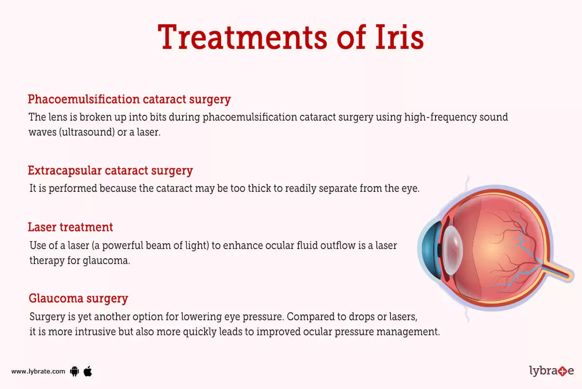
- Phacoemulsification cataract surgery: The lens is broken up into bits during phacoemulsification cataract surgery using high-frequency sound waves (ultrasound) or a laser. After removing the lens shards from your eye, the doctor inserts a new plastic lens.
- Extracapsular cataract surgery: It is performed because the cataract may be too thick to readily separate from the eye. Your ophthalmologist creates a bigger aperture in the eye during this cataract procedure. Your doctor removes the lens in one piece as opposed to first splitting it up before removing it. The produced lens is then inserted by the surgeon.
- Laser treatment: Use of a laser (a powerful beam of light) to enhance ocular fluid outflow is a laser therapy for glaucoma. Although the laser may supplement eye drops, it may not entirely replace them. The effects of laser treatments may last up to five years, although they can vary. You may repeat certain laser procedures.
- Glaucoma surgery: Surgery is yet another option for lowering eye pressure. Compared to drops or lasers, it is more intrusive but also more quickly leads to improved ocular pressure management. Although surgery may slow down visual loss, it cannot reverse it or treat glaucoma.
Iris Medicines
- Beta-adrenergic antagonists: These aid in lowering aqueous humour production. For instance, metipranolol, timolol, levobunolol, carteolol
- Prostaglandin analogues: Drugs that act as prostaglandin analogues aid in the drainage of ocular fluid. For instance, Latanoprost, Travoprost, Bimatoprost, and Tafluprost.
- Adrenergic agonists: These are drugs that encourage the aqueous humour's decreased production and increased outflow. Eg-Brimonidine
- Carbonic anhydrase inhibitors: Topical or oral drugs known as carbonic anhydrase inhibitors assist lower ocular pressure by lowering the amount of aqueous humour produced by the eye. E.g., Acetazolamide, Dorzolamide, and Brinzolamide
- Agents that are parasympathomimetic: Enhance aqueous humour discharge from the eye. often advised for open-angle glaucoma. Eg-Pilocarpine.
- Osmotic medications: These are used to treat abrupt and significant increases in intraocular pressure. In extreme circumstances, advised. E.g., Mannitol and Isosorbide.
How do I take care of my iris?
Wear sunglasses with 100% UV protection or a UV400 label anytime you’re in the sun.Tell your healthcare provider about any changes in your vision. If you wear glasses or contact lenses, have your eyes examined regularly so your provider can adjust your prescription as often as necessary.
When should I see my healthcare provider?
See your healthcare provider as soon as you notice any changes in your vision. Whether it’s something as simple as needing new glasses, or a more serious condition, don’t wait for symptoms to get worse before having your eyes checked.
Go to the emergency room if you suddenly lose your vision or have severe pain in your eyes.
Table of content


Find Ophthalmologist near me

Ask a free question
Get FREE multiple opinions from Doctors