

Pulmonary veins (Human Anatomy): Image, Functions, Diseases and Treatments
Last Updated: Mar 17, 2023
Pulmonary veins Image
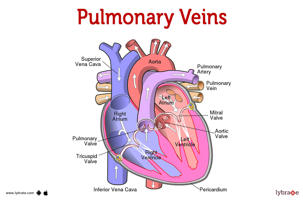
Pulmonary veins transport oxygen-rich blood to the heart. Pulmonary veins make up your pulmonary circuit. This system transports blood between the heart and lungs. These include your pulmonary arteries. The oxygen-rich blood that is produced in your lungs is collected by your pulmonary veins, which then transport it to your heart.Your pulmonary veins are susceptible to a wide variety of diseases and disorders, some of which are present from birth while others manifest themselves much later in life. Additionally, the beginnings of atrial fibrillation can be found in the pulmonary veins. As a result, people in this category are frequently the focus of treatment for A-Fib.
What is the difference between the pulmonary veins and the pulmonary arteries?
Pulmonary veins and arteries differ in two ways:
- They carry different types of blood. Your pulmonary arteries transport oxygen-depleted blood. Your pulmonary veins transport oxygenated blood.
- They travel in different directions. The pulmonary arteries transport blood from the heart to the lungs. The pulmonary veins transport blood from the lungs to the heart.
What makes pulmonary veins different from other veins?
- The only veins in the body that convey oxygen-rich blood are the pulmonary veins. All of your other veins have oxygen-deficient blood. Likewise, your pulmonary arteries are the sole arteries that transport blood deficient in oxygen. All of your other arteries transport oxygen-rich blood.
- As you can see, the pulmonary circuit is a unique part of your body! Your blood vessels in this circuit are the exceptions to the rules about the type of blood each vessel carries.
- But one aspect remains the same. That’s the direction of travel. Your pulmonary veins, like all your other veins, carry blood toward your heart. And your pulmonary arteries, like all your other arteries, carry blood away from your heart.
How many pulmonary veins are there?
Most people (60% to 70%) have four pulmonary veins. The pulmonary veins of everyone else are either three or five. There are no health problems caused by these changes in number. Some people are born with variations in their bodies that are different from what researchers consider 'normal. '
.jpg)
Where are the pulmonary veins located?
The pulmonary veins are found between the heart and the lungs. Your lungs' right and left convergent pulmonary veins are made up of several smaller blood arteries. Each pair exits its particular lung through the hilum, or root, of that lung. Your pulmonary veins continue on from that point to your heart, where they join the left atrium. Your heart's upper left chamber is seen here.
What is the structure of the pulmonary veins?
Most people have four pulmonary veins, with two connected to each lung (right and left):
- Right superior pulmonary vein: Drains your right lung’s upper lobe and middle lobe.
- Right inferior pulmonary vein: Drains your right lung’s lower lobe.
- Left superior pulmonary vein: Drains your left lung’s upper lobe and your lingula (often called the 'tongue' in your left lung).
- Left inferior pulmonary vein: Drains your left lung’s lower lobe.
- 'Superior' means above, and 'inferior' means below. Each vein's name thus gives information about where it is located and which lung it drains.
- Each pulmonary vein often has a direct connection to your left atrium. If so, your left atrium has four ostia (openings), one for each of your pulmonary veins. Through these holes, oxygen-rich blood enters your left atrium. Your blood next enters your left ventricle, where it is pumped through your aorta to your body.
How big are the pulmonary veins?
A healthy pulmonary vein has a diameter of 9 to 13 millimetres. The diameter of your pulmonary veins changes as they go from your lungs to your heart. Veins often develop broader as they go closer to your heart. This is not true for your left inferior pulmonary vein. As it emerges from your left lung, it is first broader and then becomes narrower as it approaches your heart.
What are pulmonary veins made of?
All of your veins, including your pulmonary veins, are made up of three layers of tissues and fibres. These layers are as follows:
- The tunica adventitia (outer layer): It provides your vein structure and contour.
- The tunica media (middle layer): It includes smooth muscle fibres that enable your vein to expand or contract as blood flows through it
- The tunica intima (inner layer), It has a smooth endothelial cell lining. This lining facilitates blood flow within the vein.
- Your pulmonary veins, unlike other veins in your body, are partially covered with a thin myocardial layer. This 'sleeve' of cardiac muscle tissue covers a segment of each pulmonary vein at its junction to the left atrium. This sleeve's average length is 9 millimeters. Superior pulmonary vein sleeves are longer than inferior pulmonary vein sleeves.
What anatomical variations affect the pulmonary veins?
There are several possible variations of the normal pulmonary vein structure. Some variations affect the number of pulmonary veins you have and how they enter your left atrium. These are healthy and harmless variations. Other variations prevent your pulmonary veins from draining properly into your left atrium. These variations interfere with your heart’s normal functioning and can be life-threatening.
Harmless variations
38% of persons have pulmonary vein anatomical differences that are not harmful. Their left atrium connection and the number of pulmonary veins they have are both impacted by these changes. Among these variances are:
- Common left-sided trunk. This signifies that the two left pulmonary veins join to form a single trunk that leads to the left atrium. As a result, rather of draining independently into the left atrium, the two left pulmonary veins combine before reaching it. They then empty their blood through a single hole rather than two. This popular left-sided trunk can be 'short' or 'long. ' The most frequent variety is a short left-sided trunk, which affects roughly 15% of persons.
- Accessory right pulmonary vein. This indicates that you have an additional right pulmonary vein that empties independently into the left atrium. Therefore, there are three right pulmonary veins rather than two. There are several subcategories within this primary category.
- These variances remain healthy, and your veins function normally. Because of these variances, oxygen-rich blood can still enter the left atrium. From there, the blood may travel the typical route to the left ventricle and eventually to the rest of the body.
Variations that interfere with heart function
Some variations stop your pulmonary veins from carrying oxygen-rich blood to your left atrium. So, your heart can't work as well as it should. These differences are caused by birth defects in the heart. Most of the time, they are found soon after birth, but they can also be found in adults. Among them are:
- Total anomalous pulmonary venous return (TAPVR): Children born with TAPVR have pulmonary veins that don't drain into the left atrium. Their right atrium is where their pulmonary veins really meet. The pulmonary veins of some people drain straight into their right atrium, whereas others have additional veins that go there. In either case, blood that is low in oxygen is pumped into the right side of the heart, mixing with the blood that is high in oxygen. Furthermore, their pulmonary artery is blocked off from the left side of their heart. Surgery is required for this kind of cyanotic heart disease.
- Partial anomalous pulmonary venous return (PAPVR): PAPVR is diagnosed when an infant is born with at least one of their pulmonary veins linked to their left atrium. Therefore, part of their oxygen-rich blood will be able to travel through their aorta and reach the rest of their body. These infants may still require medical attention, but in most cases, their condition is not considered to be life-threatening.
- TAPVR is detected in around one infant in every 7,809 births in the United States. There is a lack of clarity on the precise number of infants that are born with PAPVR. But according to some estimates, persons with PAPVR make up just one in every 143 people, and the condition can sometimes go undetected until maturity.
- A hole is frequently seen between the top two heart chambers of infants with TAPVR or PAPVR (atrial septal defect). Because it permits blood to flow from the baby's right atrium to their left atrium and subsequently out to their body, this defect actually saves lives. Even if this blood doesn't have as much oxygen like it should, it's still sufficient to keep the infant alive while they wait for therapy.
Pulmonary Veins Functions
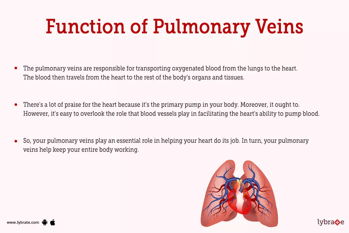
- The pulmonary veins are responsible for transporting oxygenated blood from the lungs to the heart. The blood then travels from the heart to the rest of the body's organs and tissues.
- There's a lot of praise for the heart because it's the primary pump in your body. Moreover, it ought to. However, it's easy to overlook the role that blood vessels play in facilitating the heart's ability to pump blood. Without your pulmonary veins working, your heart would not have access to oxygenated blood before pumping it to the body's other organs.
- So, your pulmonary veins play an essential role in helping your heart do its job. In turn, your pulmonary veins help keep your entire body working.
Pulmonary veins Conditions and Disorders
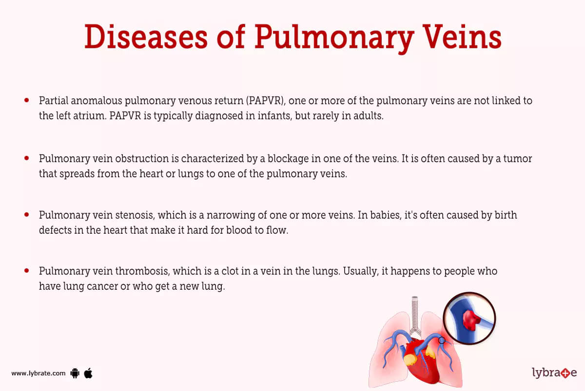
Conditions that can affect your pulmonary veins include:
- Partial anomalous pulmonary venous return (PAPVR), one or more of the pulmonary veins are not linked to the left atrium. PAPVR is typically diagnosed in infants, but rarely in adults.
- Pulmonary vein obstruction is characterized by a blockage in one of the veins. It is often caused by a tumor that spreads from the heart or lungs to one of the pulmonary veins.
- Pulmonary vein stenosis, which is a narrowing of one or more veins. In babies, it's often caused by birth defects in the heart that make it hard for blood to flow. When done to treat atrial fibrillation, catheter radiofrequency ablation can sometimes cause this condition in adults.
- Pulmonary vein thrombosis, which is a clot in a vein in the lungs. Usually, it happens to people who have lung cancer or who get a new lung.
- Pulmonary venous hypertension, which is a condition characterised by elevated blood pressure in the pulmonary veins. Normal cardiac function on the left side is frequently the culprit. Mitral valve stenosis and cardiac malignancies are two more triggers.
- Total anomalous pulmonary venous return (TAPVR), where the pulmonary veins do not communicate with the left atrium. The majority of cases with TAPVR are identified shortly after birth.
What role do the pulmonary veins play in atrial fibrillation?
Atrial fibrillation, often known as A-Fib, is an irregular cardiac rhythm that originates in the pulmonary veins of the affected individual. It's also possible that it started at the point when their atriums joined with your left one.
An atrial fibrillation episode can be caused by any of your pulmonary veins, but the most common culprit is the superior left pulmonary vein. To treat A-Fib, medical professionals use a treatment called pulmonary vein isolation, which is performed with the use of a catheter.
Pulmonary Veins Tests
- Chest X-ray: Checks for heart and lung malignancies and clots.
- CT scans: use X-rays to produce detailed images of the body. This scan looks for heart and lung cancers and structural issues.
- MR (magnetic resonance) scan: It is used to see into your organs. When it comes to finding tumours or other disorders in the veins that connect your lungs to your heart, MR scans are very helpful.
- Echocardiography: A test that visualises your heart using sound waves. This examination is performed to look for cardiac problems, such as pulmonary vein stenosis and left-sided heart failure.
Pulmonary Veins Treatments
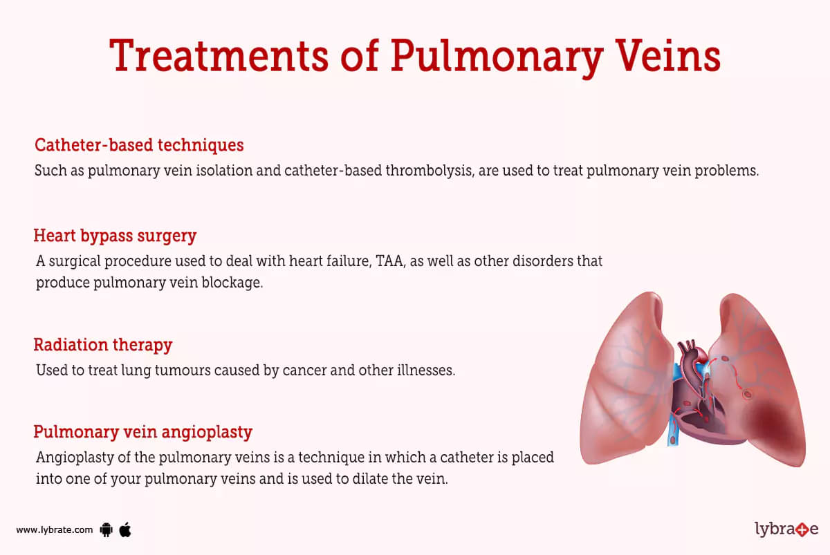
Treatments for pulmonary vein conditions include:
- Catheter-based techniques: such as pulmonary vein isolation and catheter-based thrombolysis, are used to treat pulmonary vein problems.
- Heart bypass surgery: A surgical procedure used to deal with heart failure, TAA, as well as other disorders that produce pulmonary vein blockage.
- Radiation therapy: Used to treat lung tumours caused by cancer and other illnesses.
- Pulmonary vein angioplasty: Angioplasty of the pulmonary veins is a technique in which a catheter is placed into one of your pulmonary veins and is used to dilate the vein.
- Left heart catheterization: A technique in which a long, thin tube is placed into the left ventricle (the pumping chambers of the heart) and into one of the pulmonary veins. After measuring blood pressure, temperature, as well as other health indicators, the tube is discarded.
- Sclerotherapy: A treatment option that stops the blood clots from forming by using a substance. This is used to treat pulmonary vein stenosis, which is when the walls of the pulmonary veins are too thin.
Pulmonary Veins Medicines
- Steroids for reducing inflammation of Pulmonary veins: Pain relievers and anti-inflammatory drugs. You may take them orally or inject them.
- Prednisone: An anti-inflammatory and analgesic, prednisone is a useful medication. In most cases, a daily tablet is all that is required.
- Clotting factors (fibrinogen, von Willebrand factor): These proteins contribute to the normal clotting of your blood. In order to reduce the risk of pulmonary vein thrombosis, doctors will often prescribe them in conjunction with other anticoagulants (a serious complication of heart failure).
- Analgesics for the discomfort in the pulmonary veins: Ibuprofen (Advil, Motrin), naproxen (Naprosyn), and acetaminophen (Tylenol) are examples of pain relievers that are available without a prescription and may be purchased without a doctor's visit.
- Muscle relaxants for stiffness in Pulmonary veins: Drugs to relieve stiffness and pain include baclofen (Flexeril), diazepam (Valium), and fentanyl (Duragesic).
- Antibiotics for infection in Pulmonary veins: Azithromycin is a routinely given antibiotic for pulmonary vein infections. Daily dosing is common. Doxycycline is another single-dose drug for a similar purpose.
- Nutritional supplements for reducing pain in Pulmonary veins: Several studies have demonstrated that grapeseed extract may help alleviate discomfort and inflammation in the pulmonary veins.
How can I take care of my pulmonary veins?
It's crucial to adhere to your doctor's orders if you've been identified with a problem affecting your pulmonary veins. The root cause of many pulmonary vein issues can be traced to another illness. These conditions require close monitoring and special attention throughout treatment. Consult your doctor about the best ways to handle your disease and alleviate symptoms at home.
Table of content


Find Vascular Surgeon near me

Ask a free question
Get FREE multiple opinions from Doctors