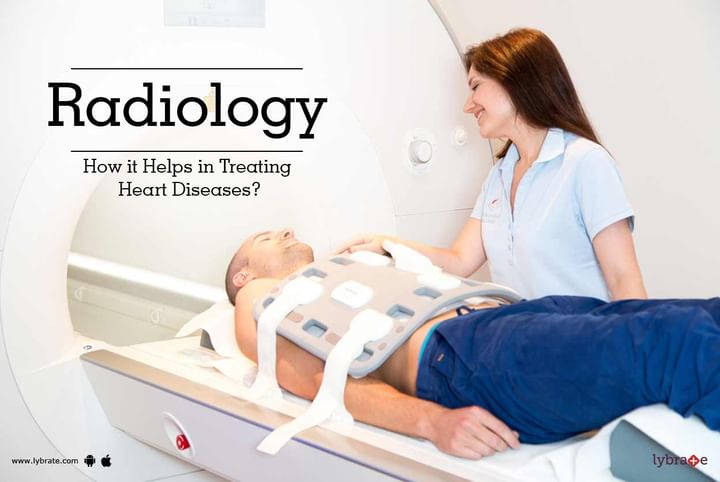
Radiology - How it Helps in Treating Heart Diseases?
Radiology is a branch of medicine that deals with diagnosis and treatment of diseases using radiant energy. Radiology uses the imaging technology like ultrasound, x-ray radiography, Magnetic Resonance Imaging (MRI), Computed Tomography (CT), Positron Emission Tomography (PET), nuclear medicine. The imaging technology helps the doctor (or physician) to see within the human body and helps you get diagnosed and treated in a better way.
Radiology is also referred to as radioscopy or clinical radiology where the latter refers to the diagnosis and treatment of fatal injuries/diseases. Radiologist is a doctor who has specialized in radiology after completion of a formal medical course to become a certified doctor. Radiographer is in turn a technician who specializes in the physical operation of scans and machines like x-rays etc. You must be well aware that the Radiologist is a professional doctor but the Radiographer is just a technician.
The following are the various uses of radiology when it comes to monitor the activity of your heart:
- MRI: It is known as Magnetic Resonance Imaging and it makes use of the energy stored in your body’s hydrogen atoms. It uses highly advanced computer 2-D and 3-D images and no ionizing radiation is used in MRI. The activity with the heart can be precisely recorded by MRI scan.
- Nuclear medicine: It involves giving your body a small tracker or a radioactive material which records the radiation coming from your body using special cameras like gamma cameras or PET. It can measure the complexity of your heart disease.
- Fluoroscopy: It helps to give real time visualization of the body by using x-rays that allow the doctor to efficiently monitor the body part, joint and bones as well as the activity that pertains to the heart.
- Ultrasound: It is a test which uses high frequency sound waves to form an image within the insides of any human organ like blood vessels or the heart. Many prefer this form of imaging test which uses sound waves instead of radiation and is considered less harmful.
- Computed Tomography: In this type of scan, a 3-D image is formed through several 2-D images taken at a single line of axis rotation. This test is useful as it gives detailed images of specific parts of the body like blood vessel, arteries, and soft tissues surrounding your heart. If you wish to discuss about any specific problem, you can consult a Radiologist.


+1.svg)
