

Talus Bone (Human Anatomy): Image, Functions, Diseases and Treatments
Last Updated: Mar 18, 2023
Talus Bone Image
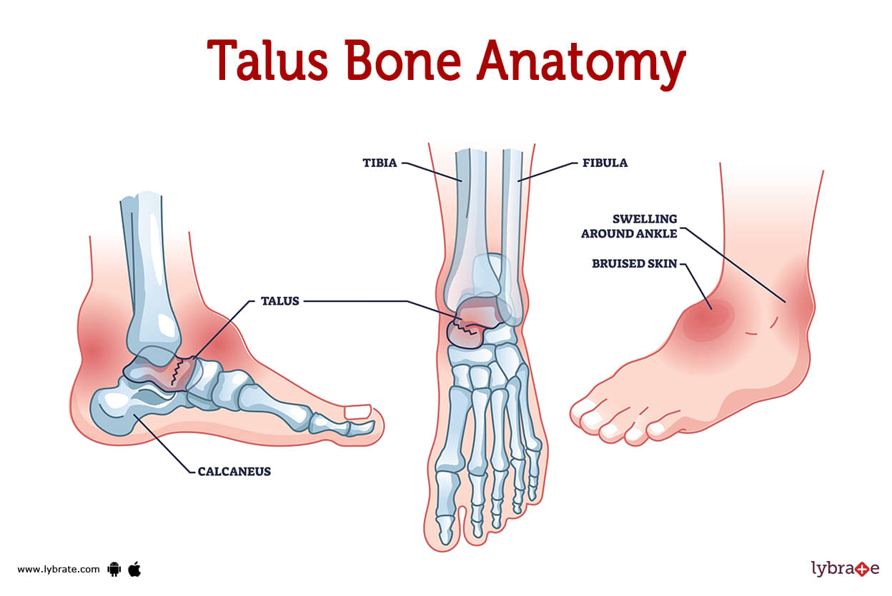
You have a little bone in your ankle called the talus. Astragalus is another name for this bone. You may find the talus, the second largest bone in your foot, at the heel (your hindfoot). The calcaneus (heel) is the only larger bone in the body.
Even though it's a small bone, your capability to stand and move depends a lot on the talus. It holds your weight and makes it easier for your ankle to move. When the talus is hurt or broken, it can take more time to heal and is more susceptible to problems than other bones. The talus, tibia (shin bone), and fibula are the three bones that come together to form your ankle joint (calf bone).
The ankle joint is formed when the talus articulates with the tibia (shin bone) and fibula (calf bone). A broken talus may necessitate surgical repair followed by physical therapy to recover mobility and strength. Osteoporosis can weaken your talus just like it does the rest of your bones.
Where is the talus located?
The talus can be found in the area of the ankle that is closer to the heel. The two bones in the lower leg, the tibia and the fibula, come together at this point to form what is known as the talus.
What does the talus look like?
A saddle-like structure can be seen in the talus. It has a domed ridge in the centre and two lower ends that are flared outward. It is coated in a covering of cartilage that performs the functions of a cushion, a shock absorber, and a lubricant, assisting in the smooth movement of your ankle.
The talus, in contrast to other bones, is not attached to any of the muscles in the body.
The talus is composed of these three parts:
- Talus head: At the top of your foot, around the navicular bone, is where the head connects to it.
- Talus body: The curving dome of the talus, which is also referred to as the trochlea of the talus, forms the ankle joint by joining the tibia and the fibula.
- Talus neck: Neck connects talus head and body. It curls down and inward.
These pieces and labels are for your doctor to utilise to indicate pain or concerns. Your doctor may use these terminology to describe a talus fracture.
How big is the talus?
The talus is not large. The length of the talus bone in the majority of adults is approximately 2 inches.
.jpg)
Talus Bone Functions
Among its many crucial functions is constructing your ankle joint, supporting your leg's weight, raising and lowering your foot, maintaining your balance (by swiping the back of your foot from side to side), stabilising the foot's arch, and helping ligaments in your foot, heel, and ankle.
Talus Bone Conditions and Disorders
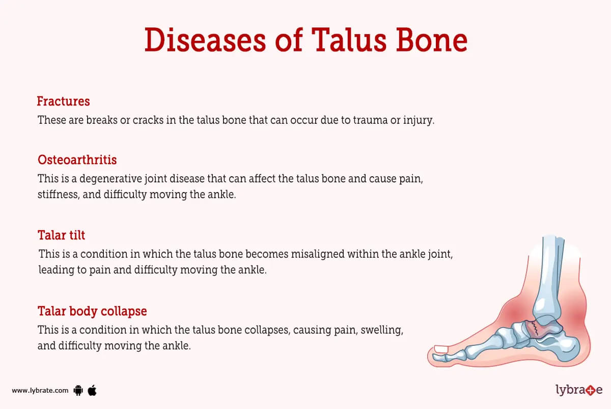
- Fractures: These are breaks or cracks in the talus bone that can occur due to trauma or injury.
- Osteoarthritis: This is a degenerative joint disease that can affect the talus bone and cause pain, stiffness, and difficulty moving the ankle.
- Talar dome lesion: This is a type of injury to the smooth, round surface of the talus bone that can cause pain and difficulty moving the ankle.
- Avascular necrosis: This is a condition in which the blood supply to the talus bone is disrupted, leading to bone death and potential collapse of the bone.
- Talar neck fracture: This is a fracture that occurs in the neck of the talus bone, which connects the talus to the tibia and fibula.
- Talar body fractures: These are fractures that occur in the main body of the talus bone, often as a result of high-impact trauma or falls.
- Talar impingement: This is a condition in which the talus bone becomes stuck or trapped in the ankle joint, leading to pain and limited range of motion.
- Talar osteochondral defects: These are small areas of damaged or missing cartilage and bone on the surface of the talus bone. They can cause pain and difficulty moving the ankle.
- Talar dome fractures: These are fractures that occur in the dome, or smooth, rounded surface, of the talus bone. They can cause pain, swelling, and difficulty moving the ankle.
- Talar tilt: This is a condition in which the talus bone becomes misaligned within the ankle joint, leading to pain and difficulty moving the ankle.
- Talar body collapse: This is a condition in which the talus bone collapses, causing pain, swelling, and difficulty moving the ankle.
- Talar dome osteonecrosis: This is a condition in which the blood supply to the dome of the talus bone is disrupted, leading to bone death and potential collapse of the bone.
- Talar subluxation: This is a condition in which the talus bone becomes partially dislocated from the ankle joint, leading to pain and difficulty moving the ankle.
- Talar dislocation: This is a condition in which the talus bone becomes completely dislocated from the ankle joint, leading to severe pain and difficulty moving the ankle.
Talus Bone Tests
- Physical examination: A medical professional will do a thorough examination of the patient's ankle and foot, including the talus bone, as part of a physical examination in order to look for any indications of injury or abnormalities. In order to evaluate the patient's mobility and range of motion, the healthcare professional may also ask the patient to move their ankle and foot in a variety of various directions.
- X-ray: This is a type of radiological imaging test that uses ionising radiation to produce images of the inside of the body. X-rays can be used to diagnose fractures, bone abnormalities, and other problems with the talus bone.
- MRI (magnetic resonance imaging): An MRI can be used to identify abnormalities with the talus bone, including fractures, osteoarthritis, and avascular necrosis, among other conditions.
- CT (computed tomography) scan: X-rays are utilised in this particular imaging technique, which produces detailed pictures of the interior of the body in the form of cross-sectional views. Fractures, anomalies of the bone, and other issues with the talus bone may all be diagnosed with the use of CT scans.
- Bone scan: This is a type of imaging test that uses a small amount of radioactive material injected into the bloodstream to produce images of the inside of the body. Bone scans can be used to diagnose problems with the talus bone, such as fractures, osteoarthritis, and avascular necrosis.
- Arthroscopy: This is a surgical procedure in which a small camera is inserted into the ankle joint through a small incision to allow the surgeon to view the inside of the joint and diagnose problems with the talus bone.
Talus Bone Treatments
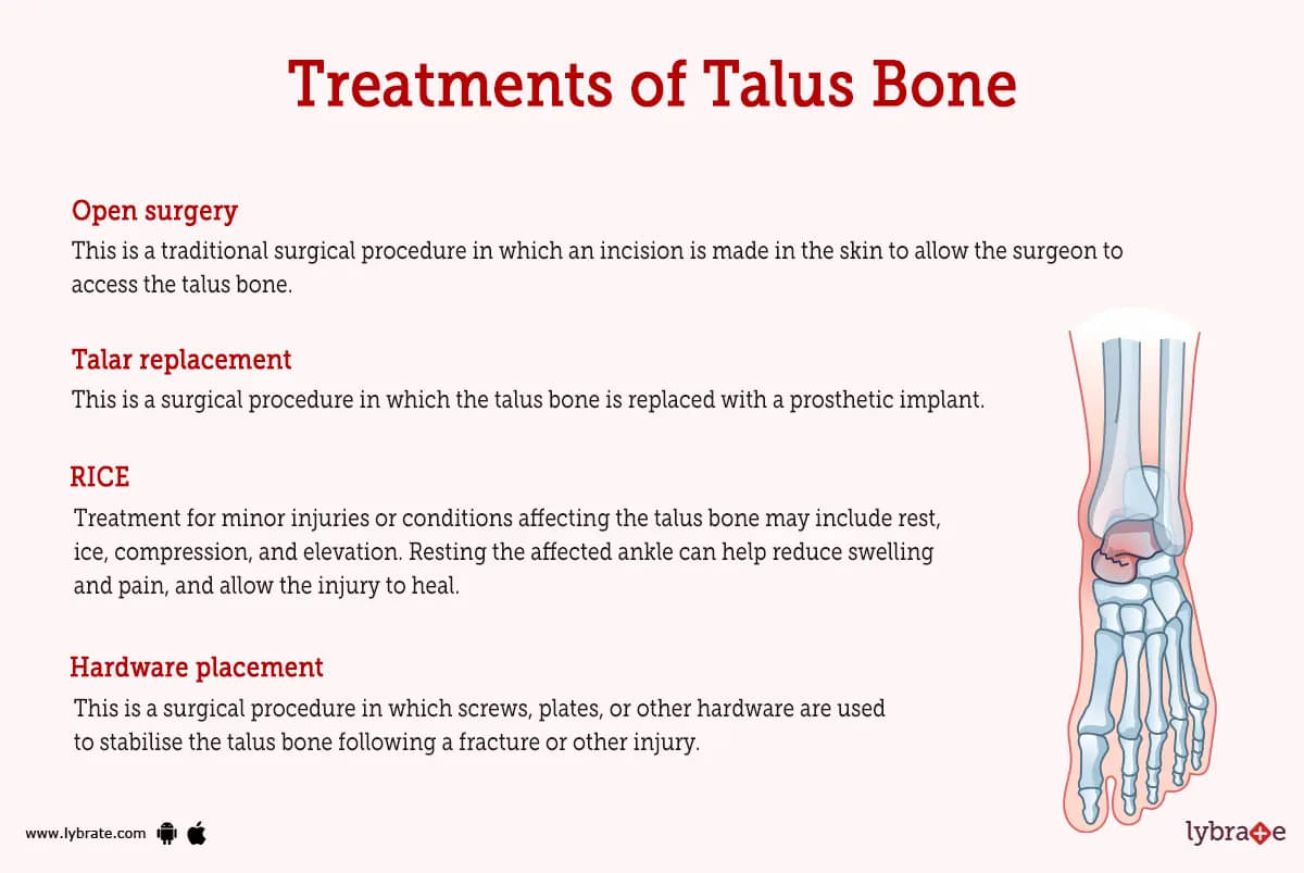
- RICE: Treatment for minor injuries or conditions affecting the talus bone may include rest, ice, compression, and elevation. Resting the affected ankle can help reduce swelling and pain, and allow the injury to heal. Applying ice to the affected ankle can help reduce swelling and pain, and it is recommended to do this for 15-20 minutes at a time, several times a day. Wrapping the affected ankle with an elastic bandage can help reduce swelling and support the ankle, and keeping the affected ankle raised above the level of the heart can help reduce swelling. It is important to consult with a healthcare professional for proper treatment and management of any injuries or conditions affecting the talus bone.
- Physical therapy: Physical therapy can help improve mobility, strength, and function of the ankle and foot, and may be recommended following an injury or surgery to the talus bone.
- Arthroscopy: This is a minimally invasive surgical procedure in which a small camera is inserted into the ankle joint through a small incision to allow the surgeon to visualise and access the inside of the joint. Arthroscopy can be used to diagnose problems with the talus bone, remove debris or loose fragments, or repair damage to the bone.
- Open surgery: This is a traditional surgical procedure in which an incision is made in the skin to allow the surgeon to access the talus bone. Open surgery may be necessary to repair fractures, remove damaged or diseased bone, or place screws or other hardware to stabilise the bone.
- Hardware placement: This is a surgical procedure in which screws, plates, or other hardware are used to stabilise the talus bone following a fracture or other injury. Hardware placement may be done using arthroscopy or open surgery.
- Osteotomy: This is a surgical procedure in which the surgeon cuts and reshapes the talus bone to improve its alignment or function within the ankle joint. Osteotomy may be necessary to correct problems such as talar tilt or talar subluxation.
- Talar replacement: This is a surgical procedure in which the talus bone is replaced with a prosthetic implant. Talar replacement may be necessary in cases of severe osteoarthritis or avascular necrosis of the talus bone.
Talus Bone Medicines
- Steroids for reducing inflammation of Talus Bone: Corticosteroids are a type of steroid that may be used to reduce inflammation in the talus bone. Some of the examples useful are prednisolone and dexamethasone.
- Analgesics for pain in Talus Bone: Ibuprofen, aspirin, and naproxen sodium are examples of typical medications that fall into this category and may be used to treat pain in the talus bone.
- Muscle relaxants for stiffness in Talus Bone: Important examples of medicines in this category may be used to treat conditions such as talar tilt or talar subluxation that cause stiffness in the talus bone are cyclobenzaprine, carisoprodol, and tizanidine
- Antibiotics for infection in Talus Bone: Amoxicillin is an example of a medicine that may be used to treat an infection in the talus bone.
- Nutritional supplements for reducing pain in Talus Bone: Some examples of supplements in this category include glucosamine and omega-3 fatty acids, which may be used to reduce pain in the talus bone.
- Supplements for promotion of growth at the time of fracture of Talus Bone: Omega-3 fatty acids, calcium, and glucosamine are examples of medicines that may be useful in promoting the growth of new bone during the healing process following a fracture of the talus bone.
Taking care of your Talus
Follow a good eating plan and exercise plan, and get regular checkups from your doctor. This will assist you in maintaining a healthy state for both your bones and your body as a whole.
If you are over the age of 50 or if osteoporosis runs in your family, you should discuss obtaining a bone density scan with your primary care physician.
If you follow these general simple guidelines, you will be less likely to get hurt:
- Always make sure you're outfitted properly for sports and other activities.
- Maintain strong bones with a healthy diet and regular exercise.
- It's important to keep your house and office free of clutter that could cause accidents.
- If you have trouble walking or are at a higher risk for falls, use a cane or walker.
- Always use your seatbelt.
- At home, you should always make use of the appropriate equipment in order to reach for objects. Keep off the furniture and the appliances.
Table of content


Find Orthopedic Doctor near me

Ask a free question
Get FREE multiple opinions from Doctors