

Vena cava (Human Anatomy): Image, Functions, Diseases and Treatments
Last Updated: Feb 02, 2023
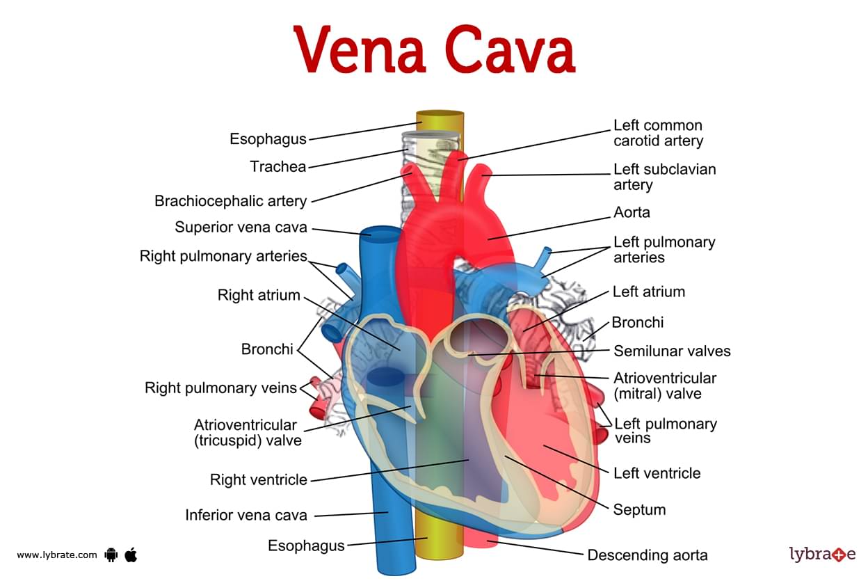
Vena cava Image
- Your superior and inferior vena cava work in tandem to return blood that has lost oxygen to your heart for fresh oxygenation. Because of this, the vena cava veins are the largest in your body. The superior vena cava receives blood from your upper body veins, and the inferior vena cava receives blood from your lower body veins.
- The superior vena cava and inferior vena cava are two of the largest veins in your body because they transport anaemic blood to your heart so that it may acquire oxygen. Blood depleted of oxygen is transported from the lower portion of your body (below your diaphragm) to your heart by the largest vein in your body, the inferior vena cava.
- Your heart receives poor quality of oxygen blood from your upper body through your superior vena cava, your second-largest vein. Think of it as a bus line. The smaller veins in your lower body that carry oxygen-depleted blood to your inferior vena cava are comparable to the downtown line (such as veins from your kidneys, liver, and lower back area).
- Blood from the other veins is transported to your heart by the inferior vena cava bus. Your superior vena cava bus carries deoxygenated blood back to your heart from your uptown line (upper body) veins, which include those in your upper back and chest. All of the deoxygenated blood that travels through the uptown and downtown bus lines eventually arrives at your heart (veins).
The vena cava's location:
- On the right side of your heart are both your inferior and superior vena cava. The right and left innominate (or brachiocephalic) veins make up the superior vena cava.
- Your right atrium, where all of your oxygen-depleted blood is sent, is where your superior vena cava, which is situated on the right side of your sternum, empties into.
- The inferior vena cava is longer than average. It begins at the junction of the right and left common iliac veins in the abdomen and ascends into the right atrium of the heart.
How does the vena cava appear?
A large vein without a valve is the superior vena cava. Your right atrium is connected to your inferior vena cava by a single valve, which is a big, long vein.
What is the size of the vena cava?
The largest veins in your body are those. Your superior vena cava measures 2 centimetres (less than 1 inch) in width and 7 centimetres (nearly 3 inches) in length. Your inferior vena cava measures around 22 millimetres, or less than 1 inch, in width and about 4 inches in length.
.jpg)
What's the composition?
Endothelial cells (which regulate nutrient exchange with tissues), Connective (supportive) tissue, Nerve fibres, Elastic fibres, and Muscle tissue all make up the superior and inferior vena cava.
Vena cava Functions
Both the superior and inferior vena cava are vitally important because they transport oxygen-depleted blood to the right atrium of the heart, from where it is pumped into the right ventricle and ultimately to the lungs via the pulmonary artery for the exchange of carbon dioxide and oxygen. Through the pulmonary veins, oxygenated blood returns to the left atrium. Blood carrying fresh oxygen then goes to your left ventricle and aorta for body distribution.
Common vena cava disorders' warning signals or symptoms
These signs may appear when anything is pressing against your superior or inferior vena cava or restricting blood flow within them. Obstruction or compression of the superior vena cava symptoms include:
- Breathing difficulty.
- Swelling in your upper body.
- Angina.
Indicators of a blood clot or tumor in the superior vena cava include:
- Edoema in the upper body
- Respiration difficulty.
Symptoms of an inferior vena cava tumor:
- Abdominal pain.
- Swelling in the legs.
- Loss of weight.
Syndrome of the inferior vena cava (obstruction or compression):
- Reduced blood pressure
- Swelling in the lower body.
- Tachycardia (rapid heart rhythm)
Vena cava Conditions and Disorders
Several conditions can affect the vena cava, including:
- Vena cava stenosis: (narrowing) occurs when the walls of the vena cava become too narrow for normal blood flow. This might occur due to an accident or the accumulation of plaque over time (the accumulation of cholesterol, fat, and other debris in the arteries).
- Aortic aneurysm: Is a bulge in the wall of the artery (an artery that carries blood rich in oxygen from the heart to the rest of the body). Aortic aneurysms can burst, which can kill you if you don't get treatment right away. Aorta dissection is a breach of the aortic wall's deepest layer. If this isn't taken care of quickly, it can cause a sudden death.
- Venous hypertension: Problems with the veins can lead to elevated blood pressure, a condition known as venous hypertension.
- Congenital malformation (something that didn't develop properly inside the mother's womb): One of the two ventricles in your brain may have a hole in its wall, a condition known as a ventricular septal defect. Seizures, unconsciousness, and even death can result from hydrocephalus (buildup of fluid on the brain).
Vena Cava Tests
- Chest X-ray for your superior vena cava: Indicates whether or not your vena cava is being compressed or obstructed in any way.
- Echocardiogram (in which a transducer is put on the chest in order to examine heart function): It is used in order to determine whether or not there is a tumour or obstruction in the superior vena cava. This can also be a sign that you are having a heart attack, as a heart attack can cause the vena cava to bulge and put pressure on the heart.
- Coronary angiography: This procedure uses a catheter and dye to visualise your superior vena cava's blood arteries. . This tests for a superior vena cava blockage or malignancy.
- Pulmonary function tests (PFTs): Used to measure lung function. These tests can detect superior vena cava blockage or malignancy.
- Ultrasound: examines veins and arteries. This is used to diagnose vena cava blood clots or malignancies.
- CT (computed tomography): takes photos of your body using x-rays. This is occasionally done to check for tumors or obstructions in the superior vena cava.
- Magnetic resonance imaging: or MRI uses radio waves and strong magnets to take pictures of your body. . This is occasionally done to check for tumors or obstructions in the superior vena cava.
- Contrast venography or phlebography (rarely used X-ray of the veins): uses a dye to check for tumours or obstructions in the vena cava.
Vena cava Treatments
There are a few therapies available for venacava issues:
- Thrombolysis of the veins (Fibrinogen and other blood-clotting agents are applied to the vein): This medication is used to disintegrate a blood clot in the vena cava.
- Angioplasty (balloon puncture of a vein to open it up and enlarge its diameter): This procedure is used to open up a blockage or tumor in your vena cava.
- Endovascular therapy (treatment using small devices inserted into the bloodstream outside of the body): Used to treat obstructions or tumours in the superior vena cava.
- Transcatheter aortic valvuloplasty (TAVP): A comparatively new procedure in which a catheter is used to open a blockage or tumor in the superior vena cava.
- Vena cava filter: The vena cava filter is a device that is put into the vena cava and collects trash and other material that might build up in the vena cava to prevent it from narrowing. Your doctor can use the same treatments for difficulties with your superior vena cava and your inferior vena cava.
- Catheter for vena cana: A catheter is put into the vena cava to get information about blood pressure as well as other health issues.
- Vena cava stent: A device put into the vena cava to aid in maintaining its patency.
- Bypass surgery for a blood clot or tumor: A procedure in which a tiny incision is made in the chest to eliminate the growth or impediment. To do this, surgeons may resort to open surgery (including a blade and the removal of tissue), minimally invasive surgery (using smaller incisions), or robot-assisted surgery (the use of imaging and computational methods in surgery).
Vena cava Medicines
- Steroids for reducing inflammation of Vena cava: prednisolone (Deltasone, Orasone, Ascriptin, Novartis Pharmaceuticals Corp. , Fort Worth, TX)
- Analgesics for pain in Vena cava: acetaminophen (Tylenol, McNeil Consumer Healthcare, Fort Worth,TX)
- Muscle relaxants for stiffness in Vena cava: diazepam (Valium, Diazepam, Alprazolam, AstraZeneca Pharmaceuticals LP, Wilmington, DE)
- Anticoagulants for preventing blood clots: warfarin (Coumadin, Jantoven, Bayer Healthcare Pharmaceuticals Inc. , West Point, PA)
- Thrombolytics for breaking up a blood clot: tissue plasminogen activator/streptokinase (tPA/streptokinase)
Tips to keep your vena cava healthy
- You may take care of your vena cava in the same way that you take care of the rest of the numerous blood vessels that are found throughout your body.
- Keep Moving.
- Take adequate care of conditions including diabetes, hypertension, and high cholesterol.
- Consume a diet that is nutritious and low in saturated fat.
- Take steps to lessen your life's stress.
Table of content


Find Vascular Surgeon near me

Ask a free question
Get FREE multiple opinions from Doctors