Trachea (Human Anatomy): Image, Function, Diseases, and Treatments
Last Updated: Feb 16, 2023
Trachea Image
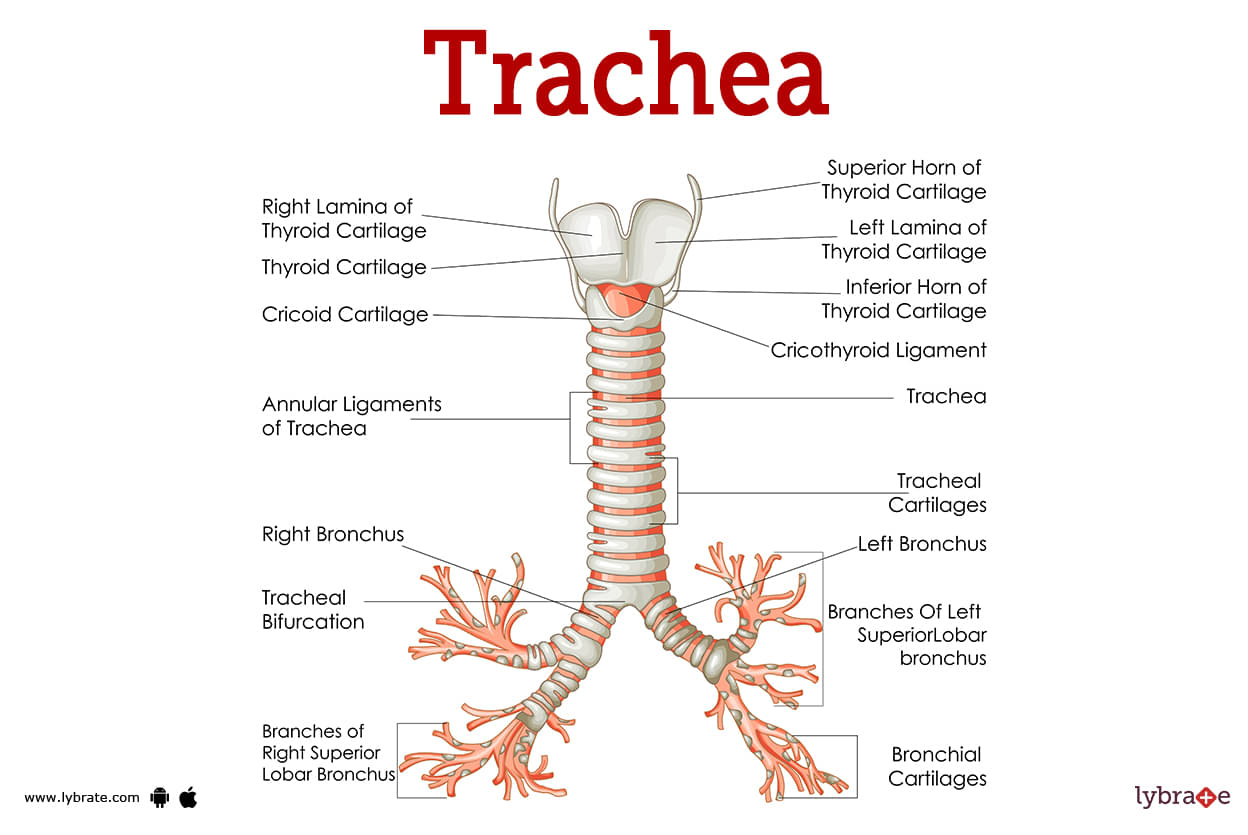
The larynx and lungs are connected by a lengthy tube called the trachea. Due to its role in sound generation, the larynx is also known as the voice box, and the trachea is often known as the windpipe.
It is a crucial component of our body's conducting and respiratory systems. Air enters our bodies via the mouth or nose, passes through the larynx, and then eventually reaches the lungs' alveoli. It belongs to the tracheobronchial tree as well.
Air enters the lungs via the tracheobronchial tree, where it exchanges gases, mostly oxygen and carbon dioxide. The trachea, bronchi, and bronchioles comprise the tracheobronchial tree. The broad tubes that connect the windpipe to the lungs are called bronchi.
The trachea is situated below the larynx, closer to the upper chest and neck. It is located between the inner borders of the collarbones, behind the notch at the bottom neck. There are two varieties of trachea. The cervical trachea, which is situated in the neck area, is the first, and the thoracic trachea, which is situated in the chest region, is the second.
Trachea Functions
The trachea's primary job is to ensure that air is transported in an orderly manner both into and out of the lungs. Because it is both rigid and flexible, the tube offers a dependable route for oxygen to enter the body. In the event that any solid, liquid, or irritant such as smoke, dust, or other particles enters the trachea, the muscles there have the ability to forcefully contract, causing coughing to remove the material. Coughing consequently serves as a kind of defence there.
Trachea Conditions
- Tracheal cancer: It is a form of cancer that begins in the windpipe and is referred to as tracheal cancer. Coughing and trouble breathing are two examples of possible symptoms. Breathing might become difficult if the trachea is compressed and narrowed by a growth such as a tumour or another development. In order to open the trachea and promote breathing, either a stent or surgery is required.
- Tracheal obstruction: A blockage in the upper airway, which might include the trachea, the larynx, or the pharynx, is referred to as a tracheal obstruction.
- Tracheal stenosis: Tracheal stenosis is a condition in which the airway becomes more constricted, which in turn makes it more difficult to breathe and may lead to a variety of complications.
- Tracheitis: Inflammation of the trachea is referred to as tracheitis and is a kind of tracheitis. The common cold or any illness that causes coughing might be the cause of this condition.
- Tracheoesophageal fistula: A fistula in the tracheoesophageal junction is an abnormal connection between the oesophagus and the trachea that may occur in one or more locations. When this condition develops, an aberrant channel arises between the trachea and the oesophagus. Serious lung conditions may result when food travels down the oesophagus and into the trachea after being eaten.
- Tracheomalacia: This condition causes the trachea to collapse in on itself. This often occurs in infants. Instead of being firm, the trachea is often flexible and floppy owing to a congenital abnormality. Tracheomalacia in adults is often brought on by trauma or smoking.
- Coughing: The body uses coughing to expel foreign objects from the lungs, trachea, and throat. Choking may happen if an item in the trachea cannot be removed. A severe instance of choking might prevent oxygen from reaching the lungs, which can result in syncope, asphyxiation, or even death.
- Stridor: The high-pitched wheezing that occurs as a result of an obstruction or constriction in the airway is referred to as stridor. It is a symptom of tracheitis that is rather prevalent.
- Laryngotracheobronchitis: Laryngotracheobronchitis is a disorder that may proceed to airway blockage and is either an inflammatory or an infectious disease.
- Vocal cord paralysis: This condition is sometimes referred to as vocal cord paralysis and vocal fold paralysis. It is a disease of the voice that manifests itself when either one or both of the vocal folds do not open or shut in the appropriate manner. A frequent kind of vocal dysfunction is called single vocal fold paralysis. It is very unusual, but paralysis of both vocal folds may be potentially fatal.
- Vascular ring anomaly: Vascular ring abnormality is an extremely uncommon kind of congenital heart malformation that occurs when the aorta develops around other organs, including the trachea and the oesophagus. In most cases, the disease becomes apparent very early in a person's life. As a result of the aorta's ability to apply pressure on both the trachea and the oesophagus, the disorder may create difficulty with breathing and eating in youngsters.
- Complete tracheal ring: A typical trachea is made up of many incomplete cartilage rings in the form of Cs. Complete tracheal rings are created when these C-shaped rings fully encircle one another to form a circle of cartilage. The youngster may have breathing difficulties due to the constriction of the windpipe caused by tracheal rings. Congenital in nature, the problem is often discovered very soon after delivery.
- Congenital high airway obstruction syndrome: A uncommon disorder known as congenital high airway obstruction syndrome results from a blockage of the baby's upper airway. This is a serious, perhaps fatal disorder that might affect the larynx or trachea tube. Due to a web-like membrane, stenosis, constriction of the airway, or lack of the trachea or larynx (tracheal or laryngeal atresia), the obstruction may be wholly or partially present.
Trachea Test
- Spirometry: Spirometry is a common medical office test that evaluates lung health by measuring inhaled air volume, exhaled air volume, and expiratory flow rate. Asthma, chronic obstructive pulmonary disease (COPD), and other respiratory illnesses can be diagnosed by spirometry.
- Lung plethysmography: It's also called body plethysmography or pulmonary plethysmography, depending on where it's being used. It helps doctors determine if their patients with lung disease have lost enough lung capacity to warrant further treatment (TLC). Total lung capacity, or TLC, is the volume of air that can be exhaled following a full inhalation.
- Flexible bronchoscopy: An endoscope is a flexible tube with a camera and light on one end that is inserted into the trachea via the mouth or nose. A professional may examine the trachea and its branches using bronchoscopy.
- Rigid bronchoscopy: The trachea is entered via the mouth using a rigid metal tube. Flexible bronchoscopy is often less successful than rigid bronchoscopy, although rigid bronchoscopy calls for heavy anaesthesia.
Using a sequence of X-rays taken by a CT scanner, a computer may produce comprehensive pictures of the trachea and adjacent structures.
A magnetic field and radio waves are used in an MRI scanner to produce pictures of the trachea and other adjacent structures. - Chest X-ray: A straightforward X-ray may show if the trachea is twisted to the left or right. An X-ray may be used to detect masses or foreign objects.
- Bronchoscopy: A bronchoscopy is a technique that enables a medical professional to see the lungs. It makes use of a bronchoscope, a small, illuminated tube. The tube is inserted via the nose or mouth, then it is pushed into the airways and down the throat. Certain lung disorders are diagnosed and treated with its aid.
- Bronchoalveolar lavage: A bronchoscopy is a common occasion for this procedure to be performed. Clearing the bronchoalveoli is another name for this process. In order to analyse a lung tissue sample, BAL is done. Before taking a fluid sample, the bronchoscope is used to flush the airways with a saline solution.
.jpg)
Trachea Treatment
- Continuous positive airway pressure (CPAP): When the patient breathes in via a face mask, air that is being held at a low pressure helps to keep the trachea open.
- Tracheobronchial airway stent: Using a bronchoscope, a thin expandable metal stent is inserted into the trachea. This allows the trachea to be kept open during the procedure.
- Tracheoplasty: A surgical procedure that provides structural support for a patient's weakened and floppy trachea to prevent tracheal collapse during breathing. Plastic mesh or the airway's surrounding tissue may be employed as a splint during surgery. The droopy section of trachea is then sutured to the support, keeping the airway open during the breathing cycle.
- Tracheostomy: An incision is made in the trachea, and a tube is then placed into the airway in order to remove the blockage that is preventing the patient from breathing normally.
- Bronchoscopic tracheal dilation: The trachea can be widened (stretched) by inserting a balloon or tracheal dilator through a bronchoscope (a light used to view the inside of the airway). This not only helps the patient breathe easier right away, but it also gives the thoracic surgeon a clearer picture of how severe the airway constriction actually is.
- Laser bronchoscopy: Scar tissue is burnt away with a laser beam using a bronchoscope; the operation gives short-term but rapid relief of the blockage.
- Tracheobronchial airway stent: A thin metallic expandable stent put into the airway using a bronchoscope is used to prop open the tracheal constriction.
- Tracheal resection and reconstruction: After the trachea has been resected to remove scar tissue and other obstructions, the two ends are resewn together to create a new, unobstructed airway.
Trachea Medicines
- Steroids for reducing inflammation of Trachea: Methylprednisolone, hydrocortisone, dexamethasone are examples of effective corticosteroids which are Drugs with anti-inflammatory properties work by preventing the recruitment of polymorphonuclear leukocytes (PMNs) to areas of cellular and tissue injury, hence reducing inflammation, especially in the region of trachea
- Analgesics for pain in Trachea: Analgesics such as aspirin, ibuprofen, and acetaminophen are all examples of medications that are able to provide at least some relief from the discomfort caused by inflammation of the laryngeal muscles and vocal cords .
- Muscle relaxants for stiffness in Trachea: Orphenadrine, metaxalone, methocarbamol, orphenadrine, tizanidine, and carisoprodol are some of the muscle relaxants that a specialist may give for stiffness and pain in laryngeal region.
- Antibiotics for infection in Trachea: These are the Medicines which are used to check the bacterial growth in lungs and treat those bacterial infections such as pneumonia. However, these meds are ineffective against viral lung disease . Common Medicines are Amoxicillin.
- Antivirals for treating infection of Trachea: These drugs are typically prescribed for five days for the treatment of nasal and bronchial congestion. Alternatively, one dose of intravenous peramivir or oral Baloxavir may be administered for one day.
- Chemotherapeutic medicines for Trachea: This type of chemotherapy comprises cyclophosphamide, doxorubicin, and 5-fluorouracil. This is followed by surgery and chest radiation treatment.
Frequently Asked Questions (FAQs)
What are the 4 functions of the trachea?
Where is your trachea?
What are the symptoms of a damaged trachea?
Can trachea be cured?
What can cause damage in trachea?
How can I keep my trachea healthy?
What is the treatment for trachea?
What diseases affect the trachea?
How do you fix your trachea?


Table of content


Find Pulmonologist near me

Ask a free question
Get FREE multiple opinions from Doctors